Supplied in Phosphate Buffered Saline, pH 7.3, with 50% Glycerol and 0.05% Proclin 300.
Storage
Shipped at 4°C. Upon delivery aliquot and store at -20°C. Avoid freeze/thaw cycles.
Synonyms
OTU domain-containing protein 7C, OTUD7C, Putative DNA-binding protein A20, TNF alpha-induced protein 3, Tumor necrosis factor alpha-induced protein 3, Zinc finger protein A20
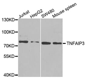
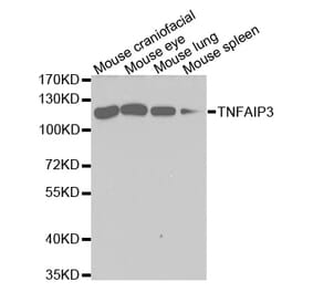
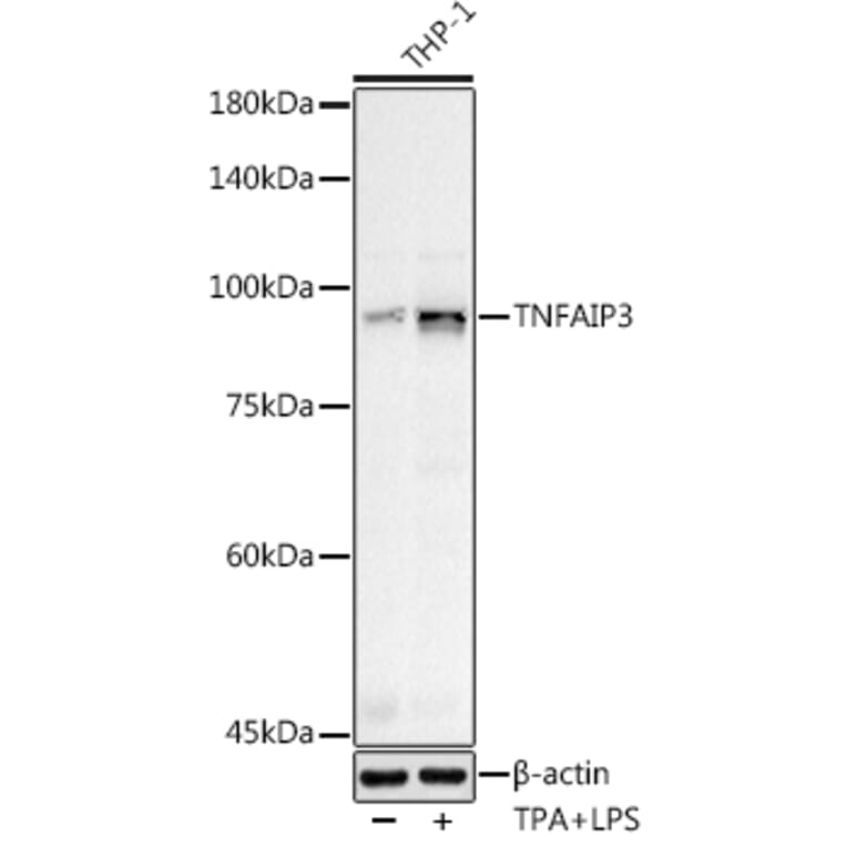
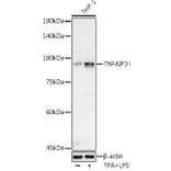
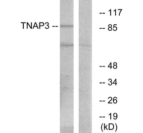
Western blot analysis of extracts of various cell lines, using Anti-TNFAIP3 Antibody (A13892) at 1:500 dilution. THP-1 cells were treated by PMA/TPA (200 nM) at 37°C for 15 minutes after serum-starvation overnight. THP-1 cells were treated by LPS (1 µg/ml) at 37°C for 8 hours. The secondary antibody was Goat Anti-Rabbit IgG H&L Antibody (HRP) at 1:10,000 dilution. Lysates/proteins were present at 25µg per lane. The blocking buffer used was 3% non-fat dry milk in TBST. Detection was with a ECL Basic Kit. Exposure time: 40s.
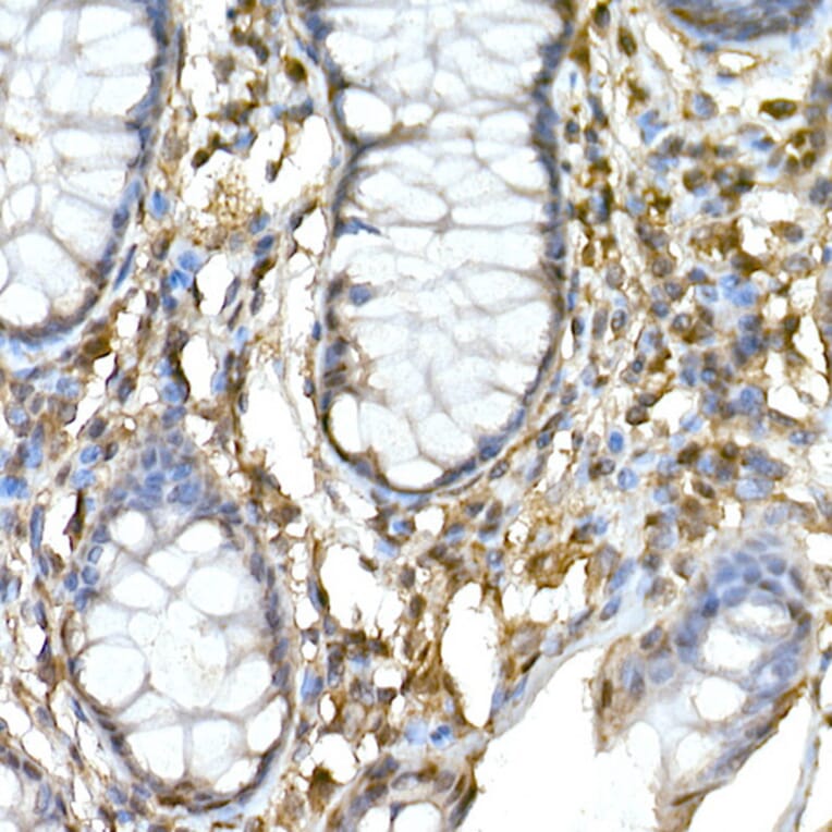
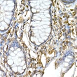
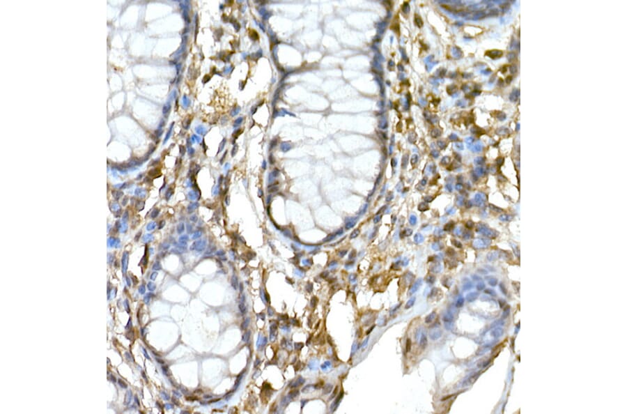
Immunohistochemistry analysis of paraffin-embedded human colon tissue using Anti-TNFAIP3 Antibody (A13892) at a dilution of 1:50 (40x lens). Perform high pressure antigen retrieval with 10 mM citrate buffer pH 6.0 before commencing with IHC staining protocol.
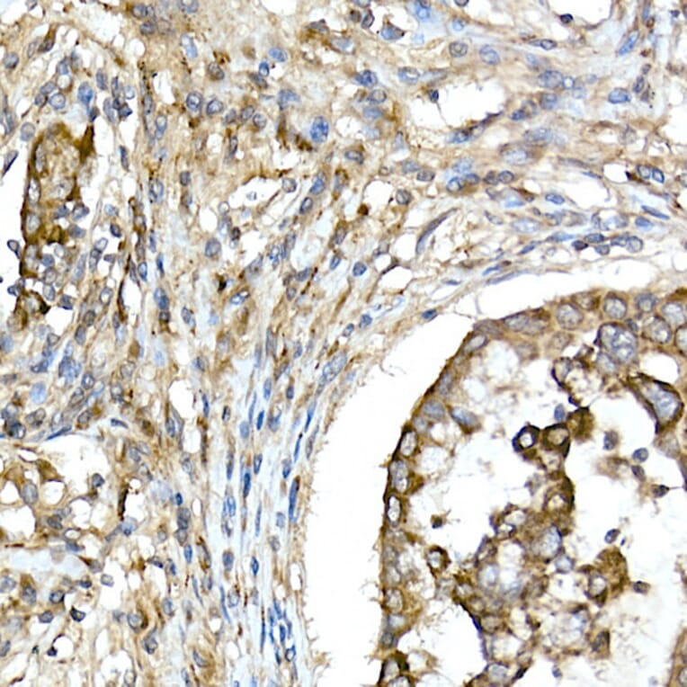
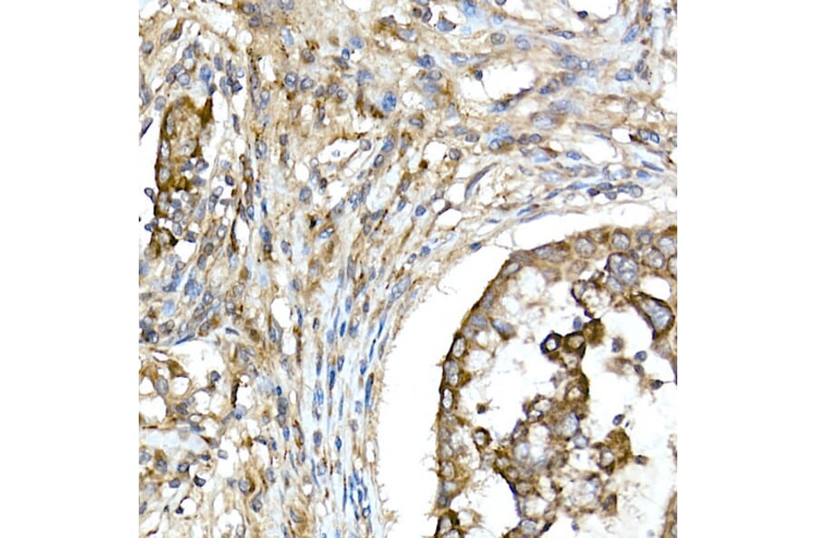
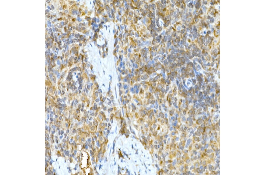
Immunohistochemistry analysis of paraffin-embedded human lung cancer using Anti-TNFAIP3 Antibody (A13892) at a dilution of 1:50 (40x lens). Perform high pressure antigen retrieval with 10 mM citrate buffer pH 6.0 before commencing with IHC staining protocol.
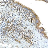
Immunohistochemistry analysis of paraffin-embedded rat lung using Anti-TNFAIP3 Antibody (A13892) at a dilution of 1:50 (40x lens). Perform high pressure antigen retrieval with 10 mM citrate buffer pH 6.0 before commencing with IHC staining protocol.
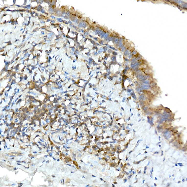
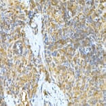
Immunohistochemistry analysis of paraffin-embedded rat spleen using Anti-TNFAIP3 Antibody (A13892) at a dilution of 1:50 (40x lens). Perform high pressure antigen retrieval with 10 mM citrate buffer pH 6.0 before commencing with IHC staining protocol.


![Western Blot - Anti-TNFAIP3 Antibody [ARC0355] (A308131) - Antibodies.com](https://cdn.antibodies.com/image/catalog/308/A308131_1.jpg?profile=product_alternative)
![Immunohistochemistry - Anti-TNFAIP3 Antibody [TNFAIP3/2813] (A250158) - Antibodies.com](https://cdn.antibodies.com/image/catalog/250/A250158_1.jpg?profile=product_alternative)
![Immunohistochemistry - Anti-TNFAIP3 Antibody [TNFAIP3/2813] - BSA and Azide free (A253338) - Antibodies.com](https://cdn.antibodies.com/image/catalog/253/A253338_1.jpg?profile=product_alternative)