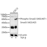
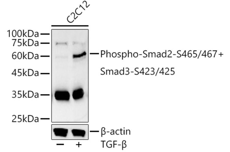
Western Blot - Anti-Smad2 (phospho Ser465 + Ser467) + Smad3 (phospho Ser423 + Ser425) Antibody (A11026)
Western blot analysis of C2C12, using Anti-Smad2 (phospho Ser465 + Ser467) + Smad3 (phospho Ser423 + Ser425) Antibody (A11026) at 1:800 dilution. C2C12 cells were treated by TGF-beta (10 ng/ml) at 37°C for 30 minutes. The secondary antibody was Goat Anti-Rabbit IgG H&L Antibody (HRP) at 1:10,000 dilution. Lysates/proteins were present at 25µg per lane. The blocking buffer used was 3% non-fat dry milk in TBST. Detection was with a ECL Basic Kit. Exposure time: 30s.
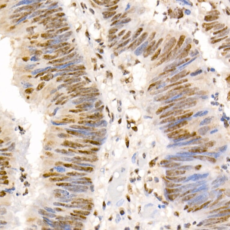
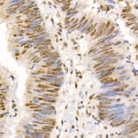
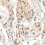
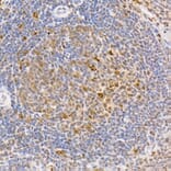
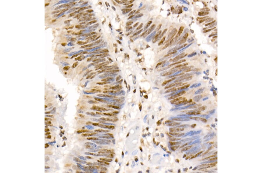
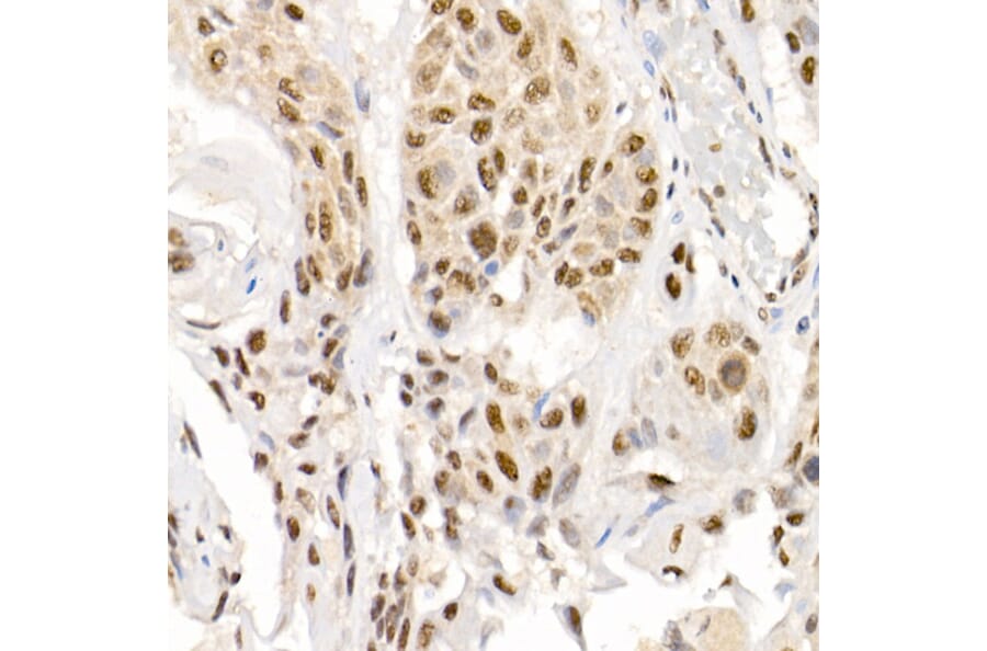
Immunohistochemistry analysis of paraffin-embedded human colon carcinoma tissue using Anti-Smad2 (phospho Ser465 + Ser467) + Smad3 (phospho Ser423 + Ser425) Antibody (A11026) at a dilution of 1:50 (40x lens). Perform high pressure antigen retrieval with 10 mM citrate buffer pH 6.0 before commencing with IHC staining protocol.
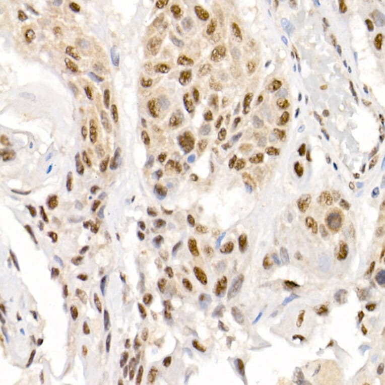
Immunohistochemistry analysis of paraffin-embedded human esophageal cancer using Anti-Smad2 (phospho Ser465 + Ser467) + Smad3 (phospho Ser423 + Ser425) Antibody (A11026) at a dilution of 1:50 (40x lens). Perform high pressure antigen retrieval with 10 mM citrate buffer pH 6.0 before commencing with IHC staining protocol.
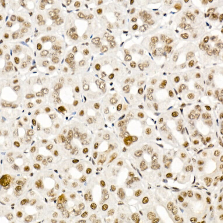
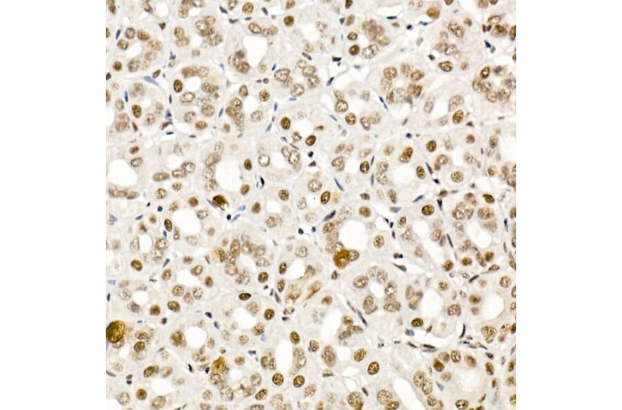
Immunohistochemistry analysis of paraffin-embedded mouse stomach using Anti-Smad2 (phospho Ser465 + Ser467) + Smad3 (phospho Ser423 + Ser425) Antibody (A11026) at a dilution of 1:50 (40x lens). Perform high pressure antigen retrieval with 10 mM citrate buffer pH 6.0 before commencing with IHC staining protocol.
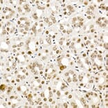
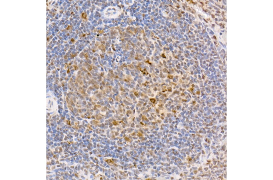
Immunohistochemistry analysis of paraffin-embedded rat spleen using Anti-Smad2 (phospho Ser465 + Ser467) + Smad3 (phospho Ser423 + Ser425) Antibody (A11026) at a dilution of 1:50 (40x lens). Perform high pressure antigen retrieval with 10 mM citrate buffer pH 6.0 before commencing with IHC staining protocol.
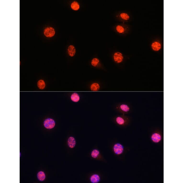
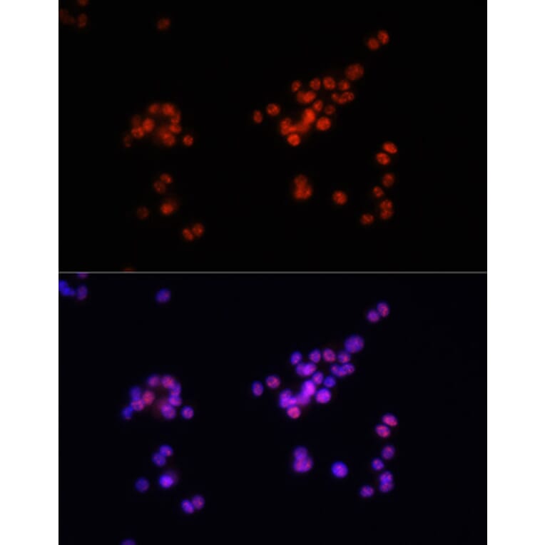
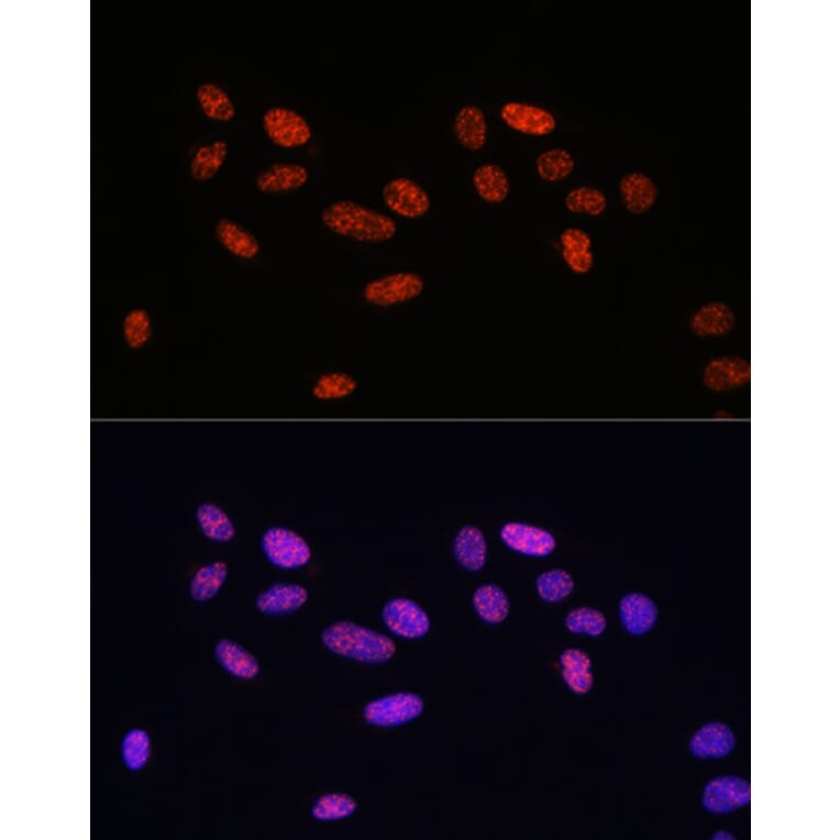
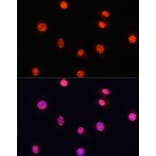
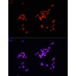
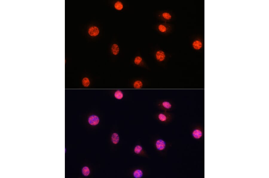
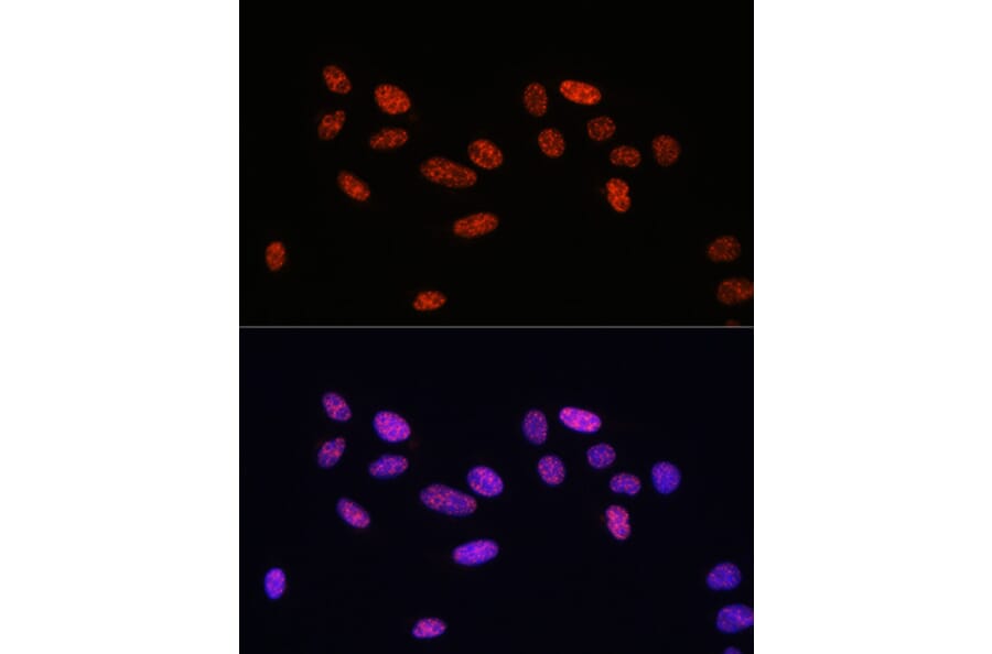
Immunofluorescence analysis of L929 cells using Anti-Smad2 (phospho Ser465 + Ser467) + Smad3 (phospho Ser423 + Ser425) Antibody (A11026) at a dilution of 1:100. DAPI was used to stain the cell nuclei (blue).
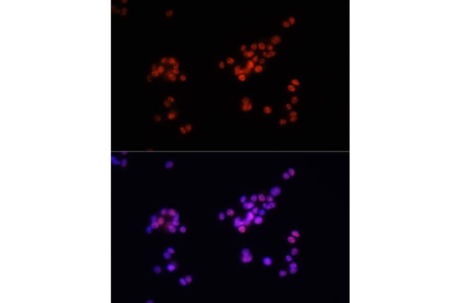
Immunofluorescence analysis of PC-12 cells using Anti-Smad2 (phospho Ser465 + Ser467) + Smad3 (phospho Ser423 + Ser425) Antibody (A11026) at a dilution of 1:100. DAPI was used to stain the cell nuclei (blue).
Immunofluorescence analysis of U2OS cells using Anti-Smad2 (phospho Ser465 + Ser467) + Smad3 (phospho Ser423 + Ser425) Antibody (A11026) at a dilution of 1:100. DAPI was used to stain the cell nuclei (blue).