Synthetic peptide within amino acids 500 and the C-terminus of human RbBP5 (NP_005048. 2).
Host
Rabbit
Clonality
Polyclonal
Isotype
IgG
Conjugate
Unconjugated
Purification
Antigen affinity purification.
Concentration
1 mg/ml
Product Form
Liquid
Formulation
Supplied in Tris-Citrate/Phosphate Buffer, pH 7-8, with 0.09% Sodium Azide.
Storage
Shipped at 4°C. Upon delivery aliquot and store at -20°C. Avoid freeze/thaw cycles.
General Notes
This antibody was affinity purified using the immunising peptide immobilized on solid support. Immunoglobulin concentration was determined by extinction coefficient: absorbance at 280 nm of 1. 4 equals 1.0 mg of IgG.
Synonyms
RBBP-5, RBQ3, Retinoblastoma-binding protein 5, Retinoblastoma-binding protein RBQ-3
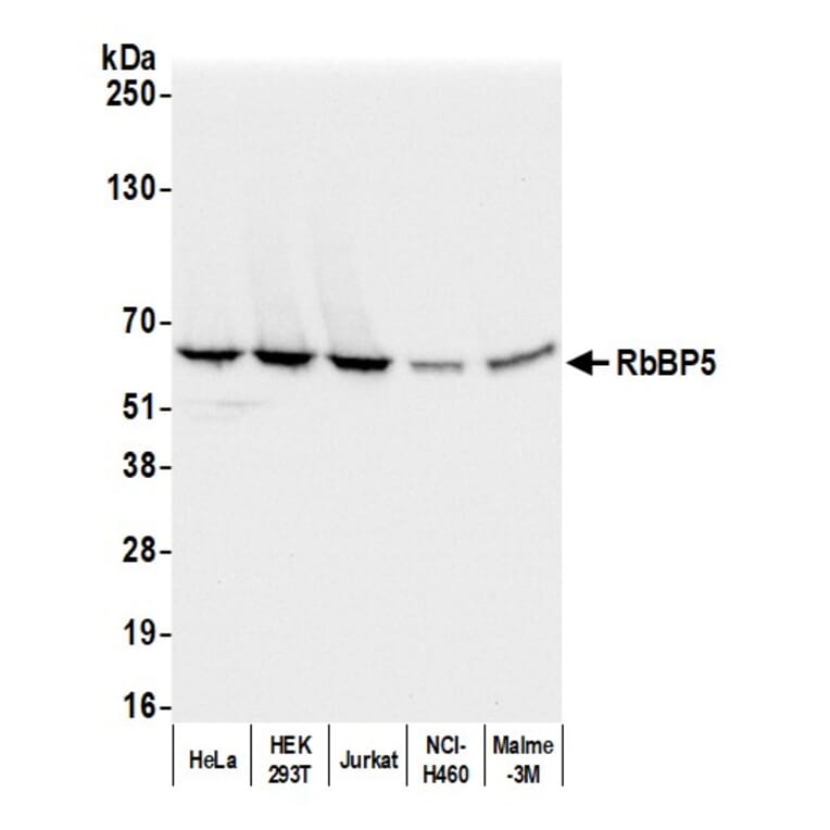
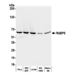
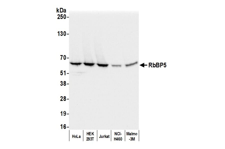
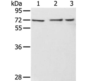
Samples: Whole cell lysate (50 µg) from HeLa, HEK293T, Jurkat, NCI-H460, and Malme-3M cells prepared using NETN lysis buffer. Antibody: Anti-RbBP5 Antibody (A295267) was used for WB at 0.04 µg/ml. Detection: Chemiluminescence with an exposure time of 3 seconds.
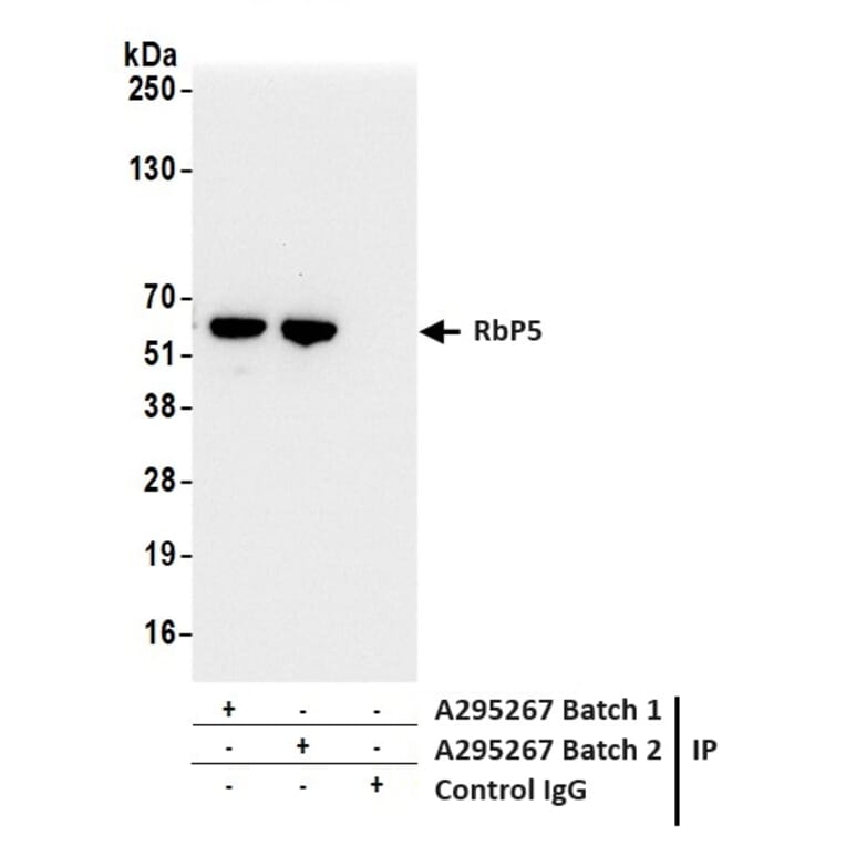
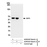
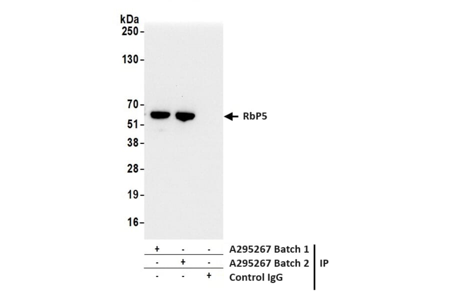
Samples: Whole cell lysate (1.0 mg per IP reaction; 20% of IP loaded) from HeLa cells prepared using NETN lysis buffer. Antibodies: Anti-RbBP5 Antibody (A295267) was used for IP at 6 µg per reaction. RbBP5 was also immunoprecipitated by a previous lot of this antibody. For blotting immunoprecipitated RbBP5, Anti-RbBP5 Antibody (A295267) was used at 0.04 µg/ml. Detection: Chemiluminescence with an exposure time of 3 seconds.
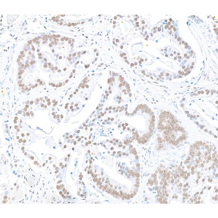
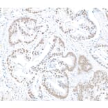
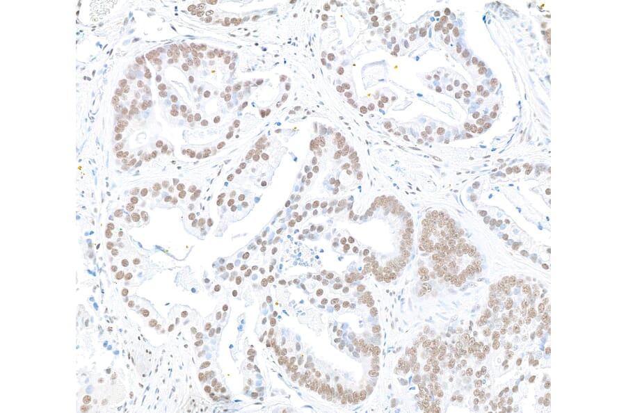
Sample: FFPE section of human prostate carcinoma. Antibody: Anti-RbBP5 Antibody (A295267) was used at a dilution of 1:5,000 (0.2 µg/ml). Detection: DAB.
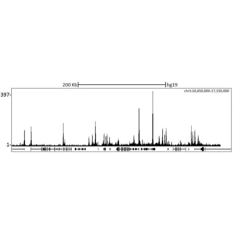
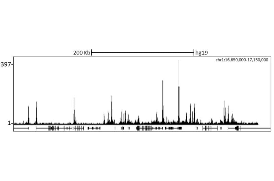
Chromatin from K562 cells was immunoprecipitated with Anti-RbBP5 Antibody (A295267) and analyzed by DNA sequencing. The figure illustrates the peak distribution of RbBP5 binding within a 500 Kb region of chromosome 1 as detected using Anti-RbBP5 Antibody (A295267). ChIP-seq validation performed by Diagenode, Denville, NJ.
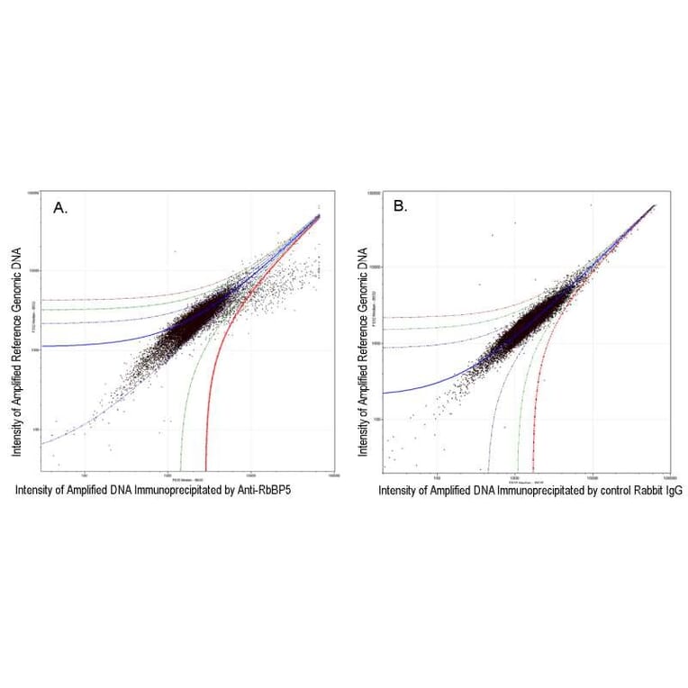
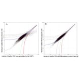
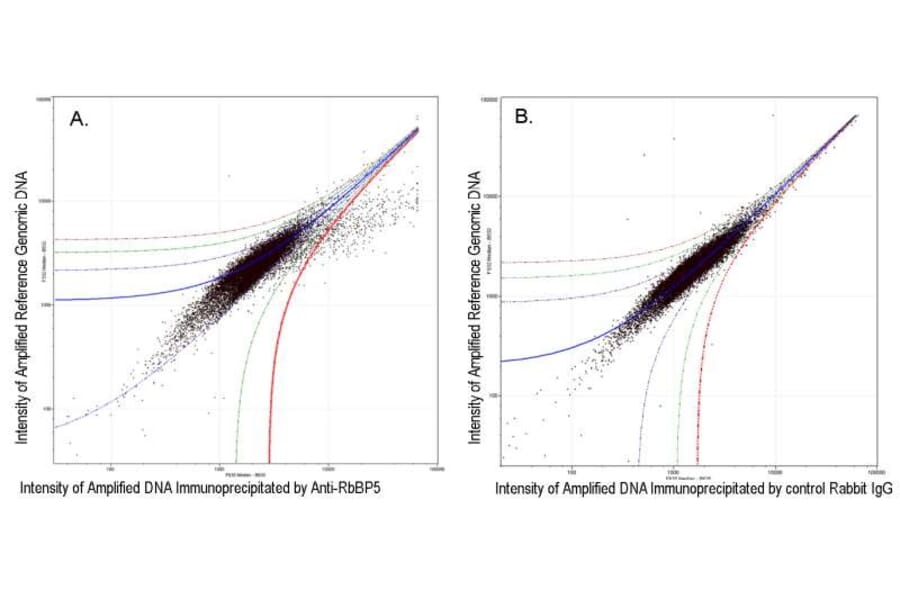
A. 10 µg of Anti-RbBP5 Antibody (A295267) was used to immunoprecipitate chromatin from K-562 cells according to Ren et al (Genes Dev. 2002 16: 245-256). Immunoprecipitated DNA and reference DNA were amplified via ligation-mediated PCR and the products labeled with fluorescent dNTPs. The labeled ChIP and reference DNA were pooled, hybridized to a DNA microarray, and analyzed. Data points below the +3 SD curve (red line) represent significantly enriched binding sites. B. As a control, a similar experiment was performed using normal rabbit IgG. Compared to the Anti-RbBP5 Antibody (A295267) ChIP, normal rabbit IgG showed little enrichment.