+1 (314) 370-6046 or
Contact Us - Argentina
- Australia
- Austria
- Bahrain
- Belgium
- Brazil
- Bulgaria
- Cameroon
- Canada
- Chile
- China
- Colombia
- Croatia
- Cyprus
- Czech Republic
- Denmark
- Ecuador
- Egypt
- Estonia
- Finland
- France
- Germany
- Greece
- Hong Kong
- Hungary
- Iceland
- India
- Indonesia
- Iran
- Ireland
- Israel
- Italy
- Japan
- Kazakhstan
- Kuwait
- Latvia
- Lithuania
- Luxembourg
- Macedonia
- Malaysia
- Malta
- Mexico
- Monaco
- Morocco
- Netherlands
- New Zealand
- Nigeria
- Norway
- Peru
- Philippines
- Poland
- Portugal
- Qatar
- Romania
- Russia
- Saudi Arabia
- Serbia
- Singapore
- Slovakia
- Slovenia
- South Africa
- South Korea
- Spain
- Sri Lanka
- Sweden
- Switzerland
- Taiwan
- Thailand
- Turkey
- Ukraine
- UAE
- United Kingdom
- United States
- Venezuela
- Vietnam

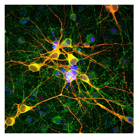
![Immunofluorescence - Anti-GAPDH Antibody [1D4] (A85382) - Antibodies.com](https://cdn.antibodies.com/image/catalog/85/A85382_1.jpg?profile=product_search)
![Flow Cytometry - Anti-PCNA Antibody [PC10] (A86877) - Antibodies.com](https://cdn.antibodies.com/image/catalog/86/A86878_948.jpg?profile=product_search)
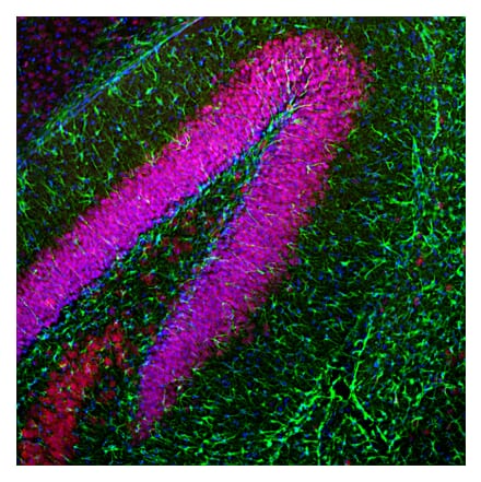

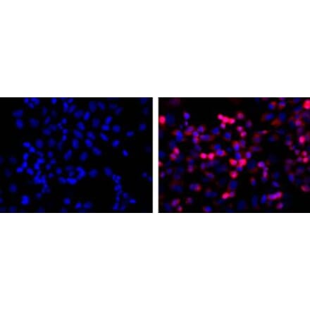
![Immunofluorescence - Anti-Fibrillarin Antibody [38F3] (A85370) - Antibodies.com](https://cdn.antibodies.com/image/catalog/85/A85370_1.jpg?profile=product_search)
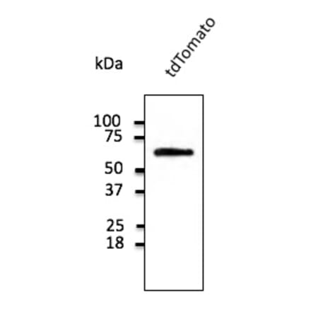
![Immunofluorescence - Anti-NeuN Antibody [1B7] (A85405) - Antibodies.com](https://cdn.antibodies.com/image/catalog/85/A85405_1.jpg?profile=product_search)
![Immunocytochemistry - Anti-beta III Tubulin Antibody [TU-20] (A86689) - Antibodies.com](https://cdn.antibodies.com/image/catalog/86/A86691_816.jpg?profile=product_search)
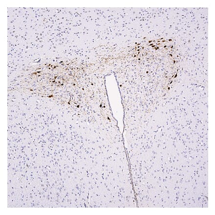
![Immunohistochemistry - Anti-mCherry Antibody [1C51] (A85305) - Antibodies.com](https://cdn.antibodies.com/image/catalog/85/A85305_1.jpg?profile=product_search)
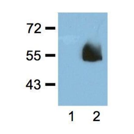
![Immunohistochemistry - Anti-Ubiquitin Antibody [Ubi-1] (A85456) - Antibodies.com](https://cdn.antibodies.com/image/catalog/85/A85456_1.jpg?profile=product_search)
![Flow Cytometry - Anti-CD107a Antibody [H4A3] - BSA and Azide free (A86536) - Antibodies.com](https://cdn.antibodies.com/image/catalog/86/A86537_711.jpg?profile=product_search)
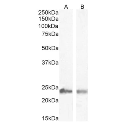
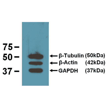
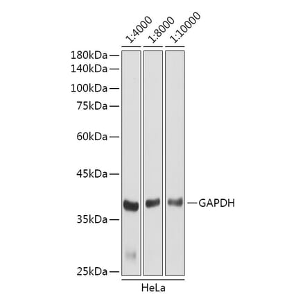
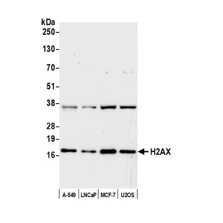
![Immunocytochemistry - Anti-alpha Tubulin Antibody [YOL1/34] (A254435) - Antibodies.com](https://cdn.antibodies.com/image/catalog/254/A254436_1.jpg?profile=product_search)
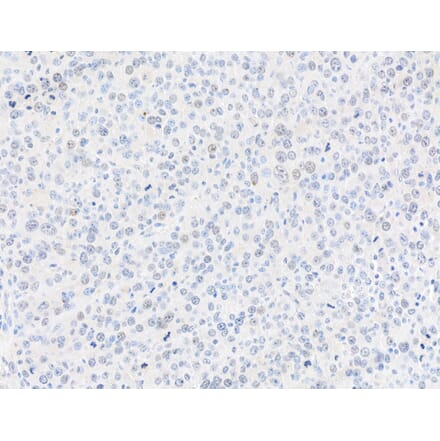
![Immunocytochemistry - Anti-Vimentin Antibody [VI-10] (A86650) - Antibodies.com](https://cdn.antibodies.com/image/catalog/86/A86652_789.jpg?profile=product_search)
![Immunohistochemistry - Anti-Cytokeratin 18 Antibody [C-04] (A86656) - Antibodies.com](https://cdn.antibodies.com/image/catalog/86/A86656_794.jpg?profile=product_search)
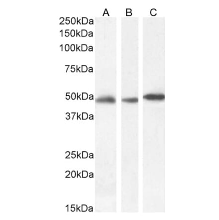
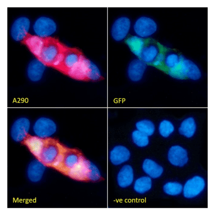
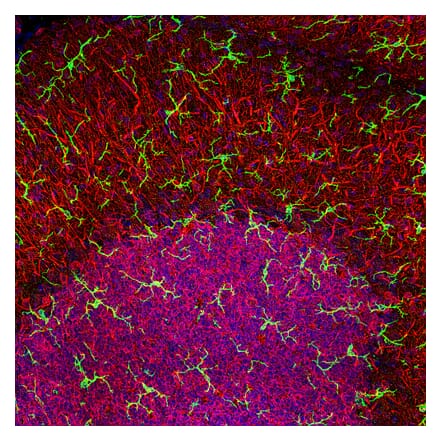
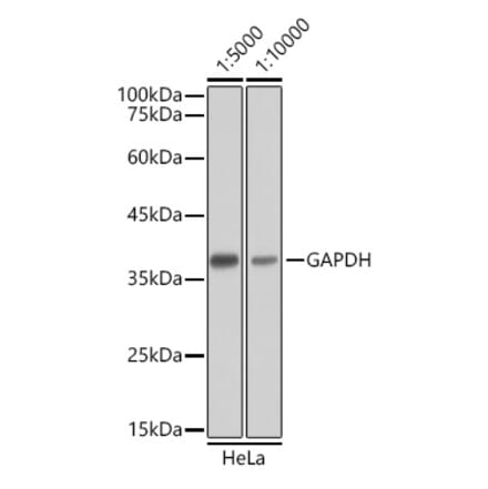
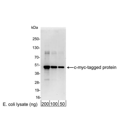
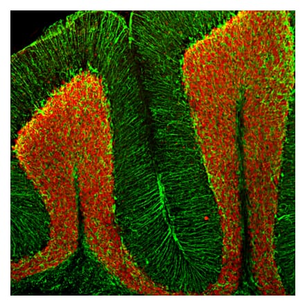
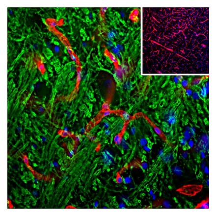
![Western Blot - Anti-DNMT1 Antibody [60B1220.1] (A304715) - Antibodies.com](https://cdn.antibodies.com/image/catalog/304/A304715_1.png?profile=product_search)
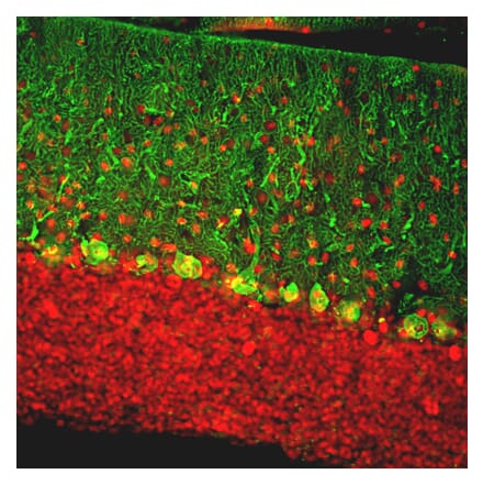
![Western Blot - Anti-LAMP2 Antibody [GL2A7] (A304947) - Antibodies.com](https://cdn.antibodies.com/image/catalog/304/A304947_1.png?profile=product_search)
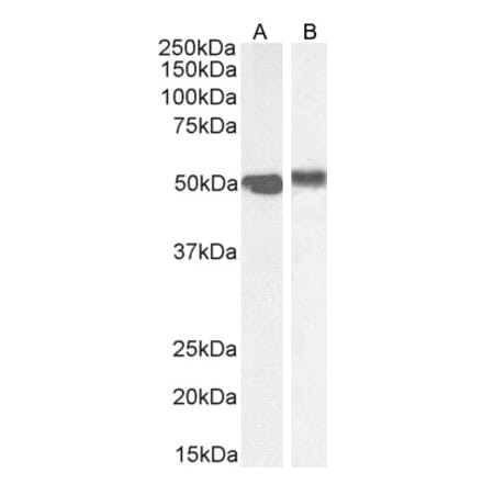
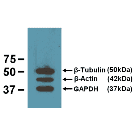
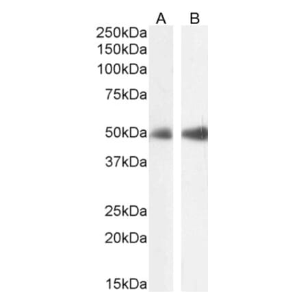
![Western Blot - Anti-Nitrotyrosine Antibody [39B6] (A304794) - Antibodies.com](https://cdn.antibodies.com/image/catalog/304/A304794_1.png?profile=product_search)
![Flow Cytometry - Anti-CD63 Antibody [MEM-259] (A85975) - Antibodies.com](https://cdn.antibodies.com/image/catalog/85/A85976_346.jpg?profile=product_search)
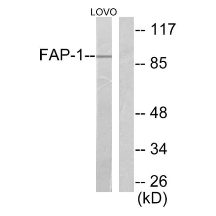
![FACS - Anti-HSP70 Antibody [C92F3A-5] (A304773) - Antibodies.com](https://cdn.antibodies.com/image/catalog/304/A304773_1.png?profile=product_search)
