+1 (314) 370-6046 or
Contact Us
  • Argentina
  • Australia
  • Austria
  • Bahrain
  • Belgium
  • Brazil
  • Bulgaria
  • Cameroon
  • Canada
  • Chile
  • China
  • Colombia
  • Croatia
  • Cyprus
  • Czech Republic
  • Denmark
  • Ecuador
  • Egypt
  • Estonia
  • Finland
  • France
  • Germany
  • Greece
  • Hong Kong
  • Hungary
  • Iceland
  • India
  • Indonesia
  • Iran
  • Ireland
  • Israel
  • Italy
  • Japan
  • Kazakhstan
  • Kuwait
  • Latvia
  • Lithuania
  • Luxembourg
  • Macedonia
  • Malaysia
  • Malta
  • Mexico
  • Monaco
  • Morocco
  • Netherlands
  • New Zealand
  • Nigeria
  • Norway
  • Peru
  • Philippines
  • Poland
  • Portugal
  • Qatar
  • Romania
  • Russia
  • Saudi Arabia
  • Serbia
  • Singapore
  • Slovakia
  • Slovenia
  • South Africa
  • South Korea
  • Spain
  • Sri Lanka
  • Sweden
  • Switzerland
  • Taiwan
  • Thailand
  • Turkey
  • Ukraine
  • UAE
  • United Kingdom
  • United States
  • Venezuela
  • Vietnam

Influenza B Virus Hemagglutinin Antibodies

Explore our collection of highly validated Influenza B Virus Hemagglutinin antibodies suitable for WB, ICC/IF, ELISA and IP that react with Influenza B Virus samples.

Showing 1-2 of 2 products
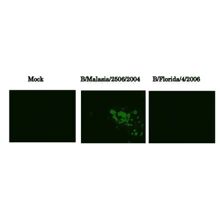
Immunofluorescence assay of MDCK (canine kidney ) cells infected with Influenza B virus, using anti-Influenza B virus HA antibody (clone 10B8). Samples were taken at 24 hours post-infection. Anti-Influenza B Virus HA antibody (clone 10B8)) efficiently detected HA in the B/Malasia/2506/2004 virus (Victorial group) but not in B/Florida/4/2006 virus (Yamagata group) infected MDCK cells. The cells were fixed with 4% paraformaldehyde in phosphate-buffered saline (PBS) and permeabilized with 0.1% Triton X-100 in PBS. The bound antibody was visualized by a further reaction with an Alexa Fluor 488-conjugated secondary antibody. Images on the left are mock-infected MDCK cells as negative control.
(2)
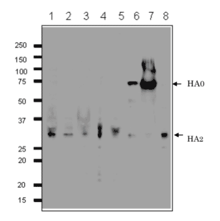
Detection of HA protein in the crude extracts of MDCK cells infected with various Influenza B virus strains by western blotting using 1H12 monoclonal antibody. 1. B/Mie/1/1993 2. B/Johannes Burg/5/1999 3. B/Florida/4/2006 4. B/Lee/1940 5. B/Florida/4/2006 6. Shandong/7/97 7. B/Malasia/2506/2004 8. B/Massachusetts/2/2012 First antibody was used at 1/500 dilution and as 2nd antibody, HRP-conjugated goat anti-mouse IgG antibody was used at 1/10,000 dilution. Positions of marker proteins are indicated in kDa on the left. Clone 1H12 recognizes an epitope on HA2 region..
(2)

Showing 1-2 of 2 products

Search Filters Contact Us 0Checkout