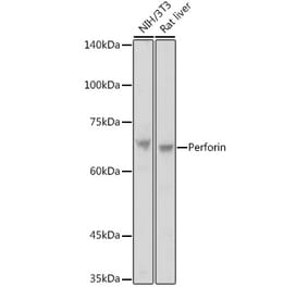
This antibody recognises perforin a 60-70 kDa glycoprotein largely expressed in CD8+ cytotoxin T lymphocytes and Natural Killer cells. It aids in defense against virus-infected or neoplastic cells by inducing apoptosis. Perforin achieves this by inserting a portion of its C-terminus region into the lipid membrane of the target in a calcium dependent manner. Subsequent polymerization with other perforin molecules forms a pore in the membrane. These pores allow the free influx and efflux of ions and proteins though the cell membrane. This disruption to homeostasis results in tonic shock which can ultimately lead to apoptosis and DNA degradation. Genetic abnormalities to the perforin gene have been shown to be linked to abnormal immune system functioning, the development of immune diseases as well as lymphomas and leukemias. This clone dG9 has been used in flow cytometry experiments to examine intracellular perforin in tumour specific CD8+ T cells incubated with autologous primary breast cancer cells and Tregs.
Applications
Flow Cytometry, IHC-Fr, IP, IHC-P, ICC
Reactivity
Human, Bovine
Cross Reactivity
This antibody does not cross react with Mouse
Immunogen
Purified granules from human YT lymphoma cell line.
Host
Mouse
Clonality
Monoclonal
Clone ID
dG9
Isotype
IgG2b
Conjugate
Unconjugated
Purification
Protein A affinity chromatography of tissue culture supernatant.
Concentration
1 mg/ml
Purity
>95% (by SDS-PAGE).
Product Form
Liquid
Formulation
Supplied in TRIS Buffered Saline with <0.1% Sodium Azide.
Storage
Store undiluted at 4°C. Do not freeze!
General Notes
Mouse anti Human perforin, clone dG9 recognizes perforin a 60-70 kDa glycoprotein largely expressed in CD8+ cytotoxin T lymphocytes and Natural Killer cells. It aids in defense against virus-infected or neoplastic cells by inducing apoptosis. Perforin achieves this by inserting a portion of its C-terminus region into the lipid membrane of the target in a calcium dependent manner. Subsequent polymerization with other perforin molecules forms a pore in the membrane. These pores allow the free influx and efflux of ions and proteins though the cell membrane. This disruption to homeostasis results in tonic shock which can ultimately lead to apoptosis and DNA degradation (Osinska et al. 2014). Genetic abnormalities to the perforin gene have been shown to be linked to abnormal immune system functioning, the development of immune diseases as well as lymphomas and leukemias (Osinska et al. 2014). This clone dG9 has been used in flow cytometry experiments to examine intracellular perforin in tumour specific CD8+ T cells incubated with autologous primary breast cancer cells and Tregs (Su et al. 2017).


![Immunohistochemistry - Anti-Perforin Antibody [PRF1/2470] (A249713) - Antibodies.com](https://cdn.antibodies.com/image/catalog/249/A249713_1.jpg?profile=product_alternative)

![Immunohistochemistry - Anti-Perforin Antibody [PRF1/2470] - BSA and Azide free (A252893) - Antibodies.com](https://cdn.antibodies.com/image/catalog/252/A252893_1.jpg?profile=product_alternative)
![Protein Array - Anti-Perforin Antibody [PRF1/2468] (A249711) - Antibodies.com](https://cdn.antibodies.com/image/catalog/249/A249712_1.jpg?profile=product_alternative)
![Protein Array - Anti-Perforin Antibody [PRF1/2468] - BSA and Azide free (A252891) - Antibodies.com](https://cdn.antibodies.com/image/catalog/252/A252892_1.jpg?profile=product_alternative)
![Immunohistochemistry - Anti-Perforin Antibody [PRF1/7077R] (A278049) - Antibodies.com](https://cdn.antibodies.com/image/catalog/278/A278049_1.jpg?profile=product_alternative)
![Immunohistochemistry - Anti-Perforin Antibody [PRF1/7077R] - BSA and Azide free (A278637) - Antibodies.com](https://cdn.antibodies.com/image/catalog/278/A278637_1.jpg?profile=product_alternative)
![Protein Array - Anti-Perforin Antibody [PRF1/2467] (A249709) - Antibodies.com](https://cdn.antibodies.com/image/catalog/249/A249710_1.jpg?profile=product_alternative)