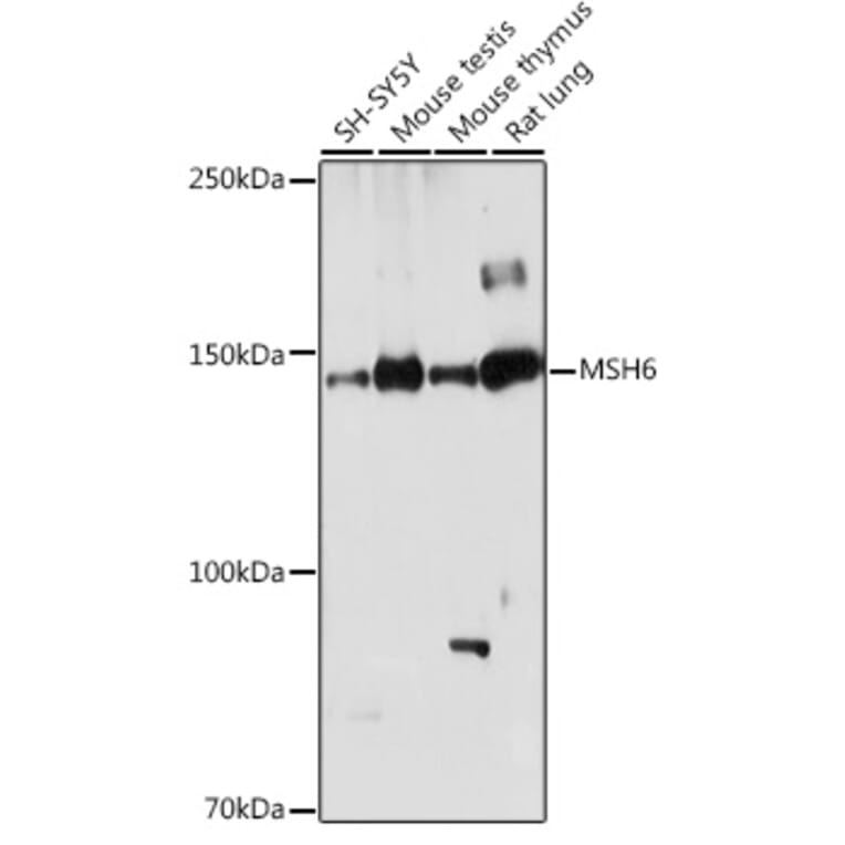
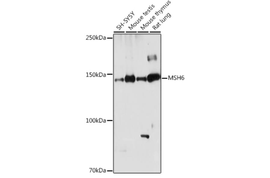
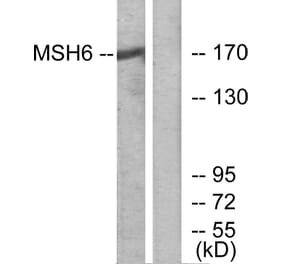
Western blot analysis of extracts of various cell lines, using Anti-MSH6 Antibody (A307834) at 1:1,000 dilution. The secondary antibody was Goat Anti-Rabbit IgG H&L Antibody (HRP) at 1:10,000 dilution. Lysates/proteins were present at 25µg per lane. The blocking buffer used was 3% non-fat dry milk in TBST. Detection was with a ECL Basic Kit. Exposure time: 180s.
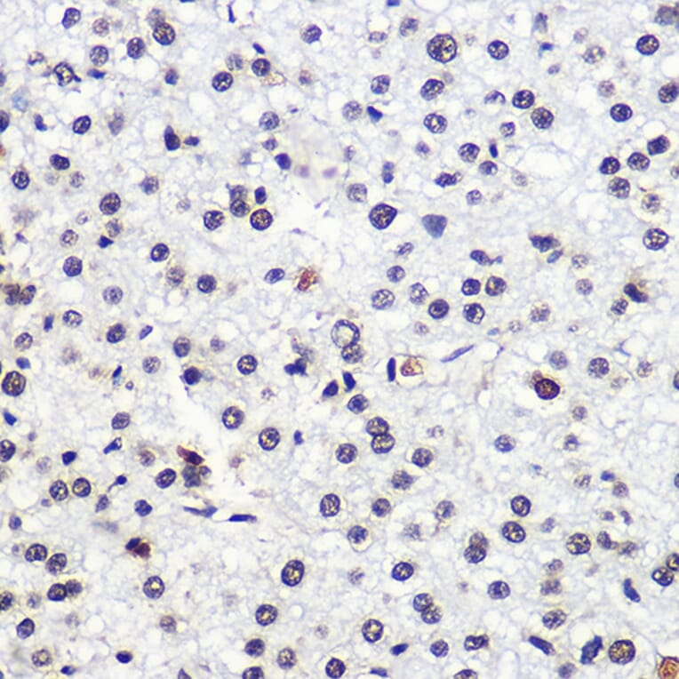
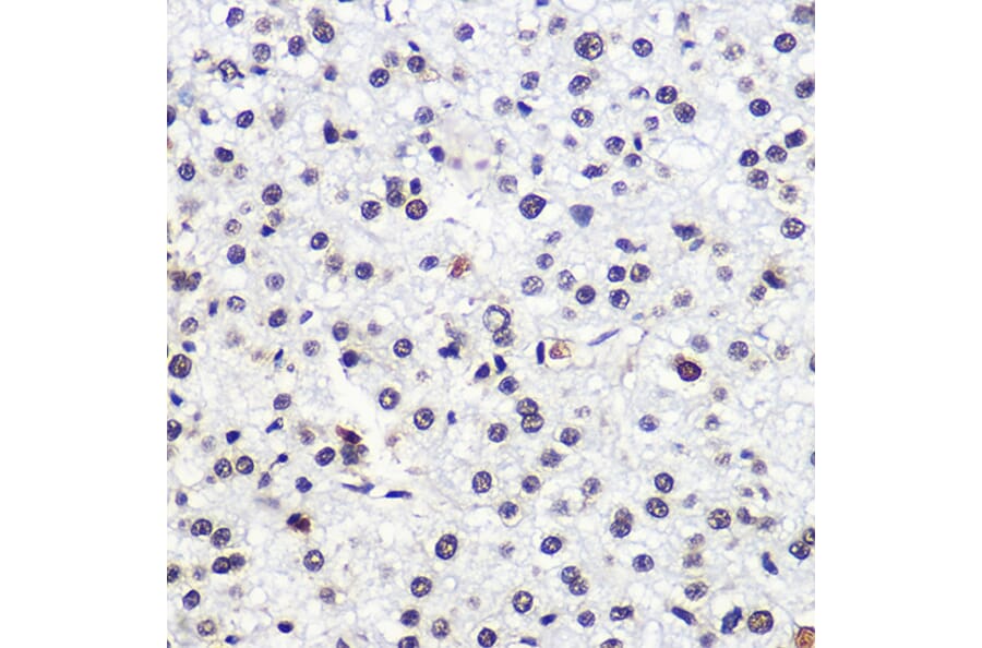
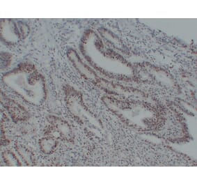
Immunohistochemistry analysis of paraffin-embedded human liver cancer using Anti-MSH6 Antibody (A307834) at a dilution of 1:100 (40x lens). Perform microwave antigen retrieval with 10 mM Tris/EDTA buffer pH 9.0 before commencing with IHC staining protocol.
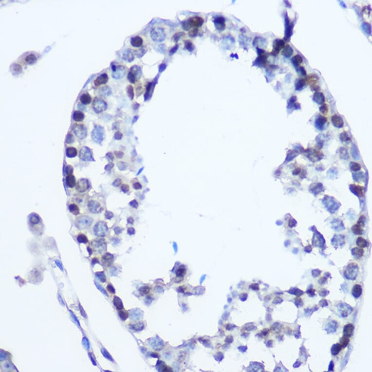
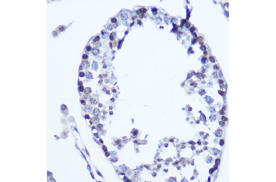
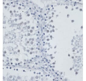
Immunohistochemistry analysis of paraffin-embedded mouse testis using Anti-MSH6 Antibody (A307834) at a dilution of 1:100 (40x lens). Perform microwave antigen retrieval with 10 mM Tris/EDTA buffer pH 9.0 before commencing with IHC staining protocol.
Immunohistochemistry analysis of paraffin-embedded rat testis using Anti-MSH6 Antibody (A307834) at a dilution of 1:100 (40x lens). Perform microwave antigen retrieval with 10 mM Tris/EDTA buffer pH 9.0 before commencing with IHC staining protocol.
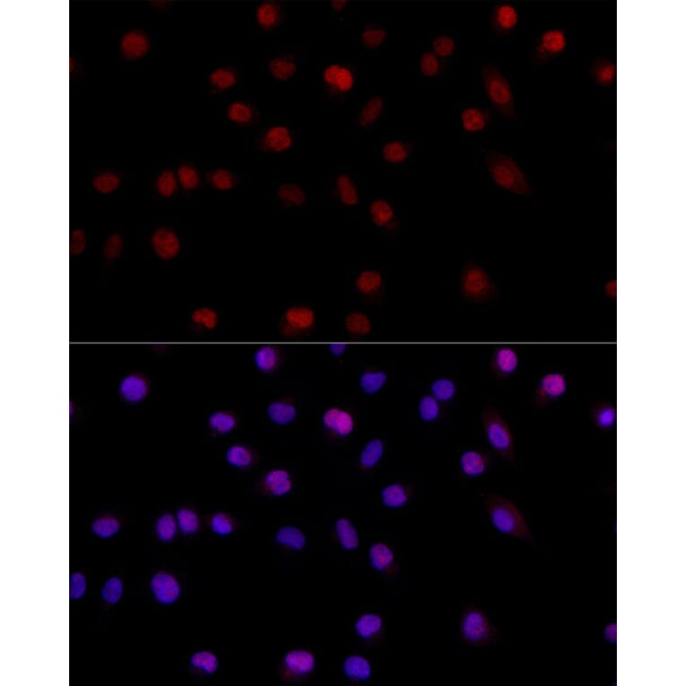
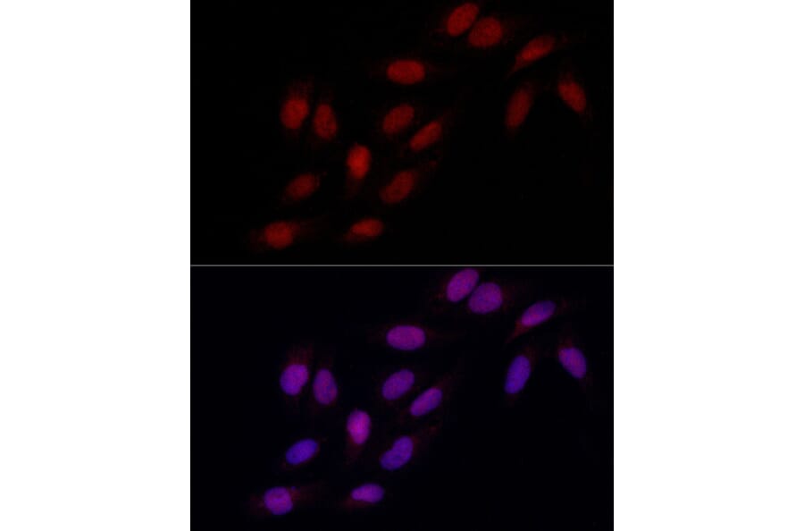
Immunofluorescence analysis of A-549 cells using Anti-MSH6 Antibody (A307834) at a dilution of 1:200 (40x lens). DAPI was used to stain the cell nuclei (blue).
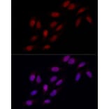
Immunofluorescence analysis of U2OS cells using Anti-MSH6 Antibody (A307834) at a dilution of 1:200 (40x lens). DAPI was used to stain the cell nuclei (blue).


![Immunohistochemistry - Anti-MSH6 Antibody [MSH6/3085] (A248784) - Antibodies.com](https://cdn.antibodies.com/image/catalog/248/A248784_1.jpg?profile=product_alternative)
![Immunohistochemistry - Anti-MSH6 Antibody [MSH6/3086] - BSA and Azide free (A251965) - Antibodies.com](https://cdn.antibodies.com/image/catalog/251/A251965_1.jpg?profile=product_alternative)
![Immunohistochemistry - Anti-MSH6 Antibody [MSH6/3085] - BSA and Azide free (A251964) - Antibodies.com](https://cdn.antibodies.com/image/catalog/251/A251964_1.jpg?profile=product_alternative)
![Immunohistochemistry - Anti-MSH6 Antibody [MSH6/3086] (A248785) - Antibodies.com](https://cdn.antibodies.com/image/catalog/248/A248785_1.jpg?profile=product_alternative)
![Immunohistochemistry - Anti-MSH6 Antibody [MSH6/3091] (A248780) - Antibodies.com](https://cdn.antibodies.com/image/catalog/248/A248780_1.jpg?profile=product_alternative)
![Immunohistochemistry - Anti-MSH6 Antibody [MSH6/3091] - BSA and Azide free (A251960) - Antibodies.com](https://cdn.antibodies.com/image/catalog/251/A251960_1.jpg?profile=product_alternative)
![Immunohistochemistry - Anti-MSH6 Antibody [MSH6/3089] - BSA and Azide free (A251966) - Antibodies.com](https://cdn.antibodies.com/image/catalog/251/A251966_1.jpg?profile=product_alternative)
![Immunohistochemistry - Anti-MSH6 Antibody [MSH6/3089] (A248786) - Antibodies.com](https://cdn.antibodies.com/image/catalog/248/A248786_1.jpg?profile=product_alternative)
![Immunohistochemistry - Anti-MSH6 Antibody [MSH6/4592R] (A277992) - Antibodies.com](https://cdn.antibodies.com/image/catalog/277/A277992_1.jpg?profile=product_alternative)