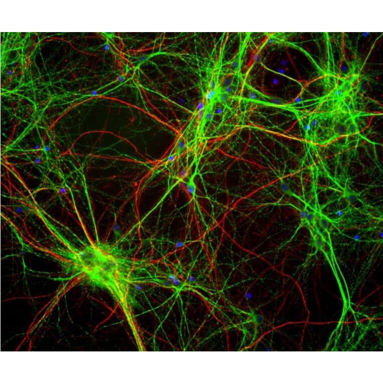
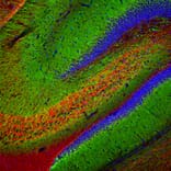
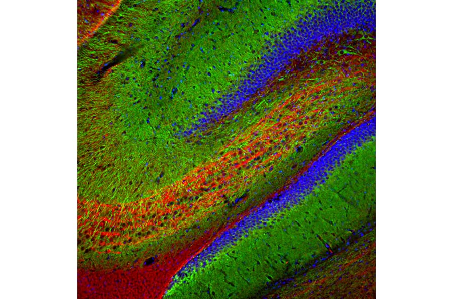
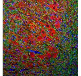
Immunofluorescent analysis of rat hippocampus section stained with Anti-MAP2 Antibody (1:5,000 | green) and Anti-a-Internexin Antibody (A85441 | 1:2,000 | red). Following transcardial perfusion of rat with 4% paraformaldehyde, brain was post fixed for 24 hours, cut to 45µM, and free-floating sections were stained with above antibodies. The Anti-MAP2 Antibody labels MAP2 protein in the perikarya and dendrites of the most neurons, and the Anti-a-Internexin Antibody selectively stains axons and dendrites of neuronal cells.
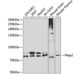
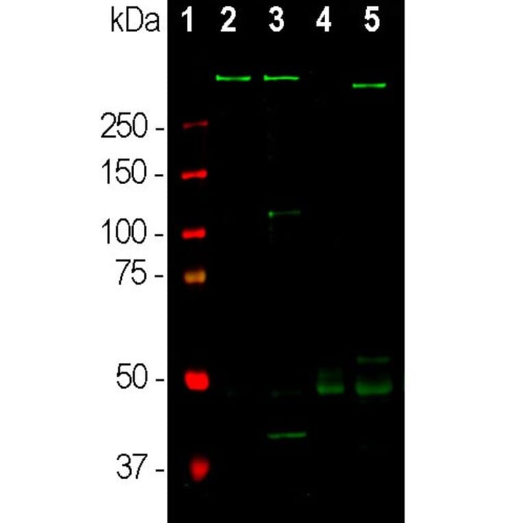
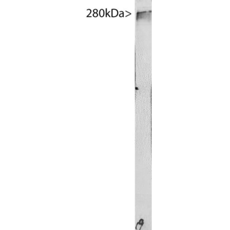
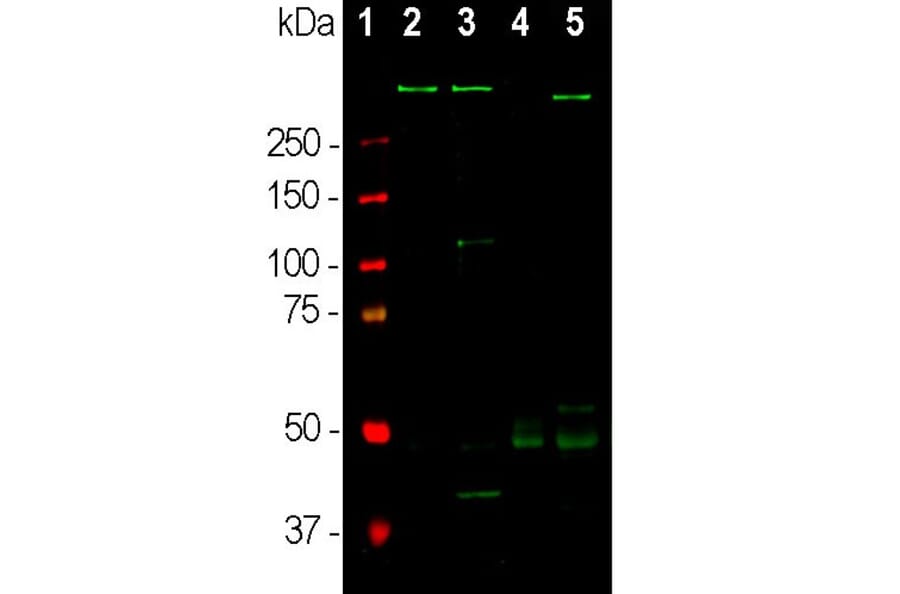
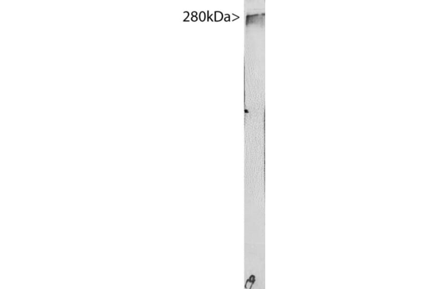
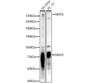
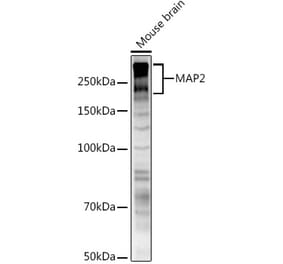
Western blot analysis of different tissue lysates using Anti-MAP2 Antibody (1:10,000 | green): [1] protein standard (red), [2] adult rat whole brain, [2] embryonic (E20) rat brain, [4] adult rat spinal cord, and [5] adult mouse brain lysate. A band at about 280 kDa corresponds to full length MAP2a and MAP2B protein. MAP2A/B is expressed heavily in brain particularly in cortical regions, but is a more minor component of spinal cord. Note that the epitope for this antibody in within the “projection domain”, and so the antibody does not bind to the lower molecular weight MAP2C and MAP2D isoforms which lack this region.
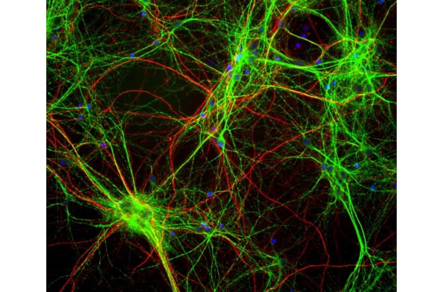
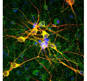
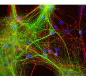
Mixed neuron/glia cultures stained with Anti-MAP2 Antibody (green) and Anti-NF-H Antibody (A85336 | red). Since the NF-H protein is largely expressed in neuronal axons, while the MAP2 is only found in neuronal dendrites and perikarya, there is little overlap between these two staining patterns. DNA stain shows nuclei of neurons and non-neuronal cells (blue).


![Immunofluorescence - Anti-MAP2 Antibody [2C4] (A85459) - Antibodies.com](https://cdn.antibodies.com/image/catalog/85/A85459_1.jpg?profile=product_alternative)
![Western Blot - Anti-MAP2 Antibody [MT-07] (A86616) - Antibodies.com](https://cdn.antibodies.com/image/catalog/86/A86618_762.jpg?profile=product_alternative)
![Western Blot - Anti-MAP2 Antibody [MT-08] (A86618) - Antibodies.com](https://cdn.antibodies.com/image/catalog/86/A86619_763.jpg?profile=product_alternative)
![Western Blot - Anti-MAP2 Antibody [MT-01] (A86785) - Antibodies.com](https://cdn.antibodies.com/image/catalog/86/A86786_893.jpg?profile=product_alternative)