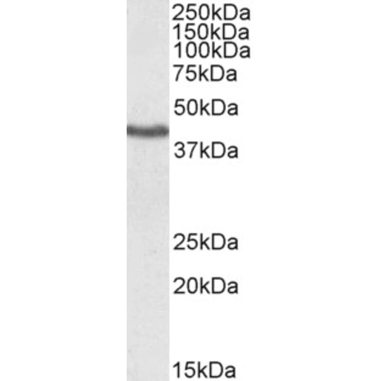
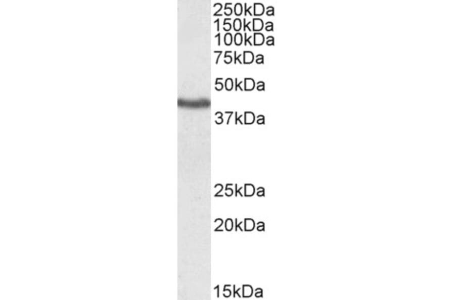
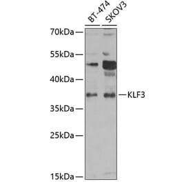
KLF3 expression in Human Heart lysate analyzed by western blot. Cells were lysed in RIPA buffer and 35µg protein was run per lane. Primary antibody incubation was performed with Anti-KLF3 Antibody (A83826) at 1µg/ml and detected by chemiluminescence.
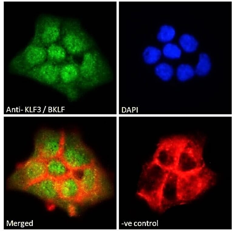
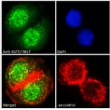
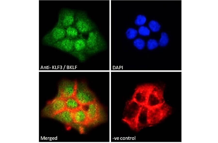
KLF3 expression in A431 cells analyzed by immunofluorescence. Cells were permeabilized with 0.15% Triton. Staining was performed with Anti-KLF3 Antibody (A83826) at 10µg/ml for 1 hour and Alexa Fluor 488 secondary antibody at 2µg/ml. Nuclear staining shown and nuclei were stained with DAPI (blue) while actin filaments were stained with phalloidin (red). Negative control: Goat IgG Isotype Control at 10µg/ml followed by Alexa Fluor 488 secondary antibody at 2µg/ml.
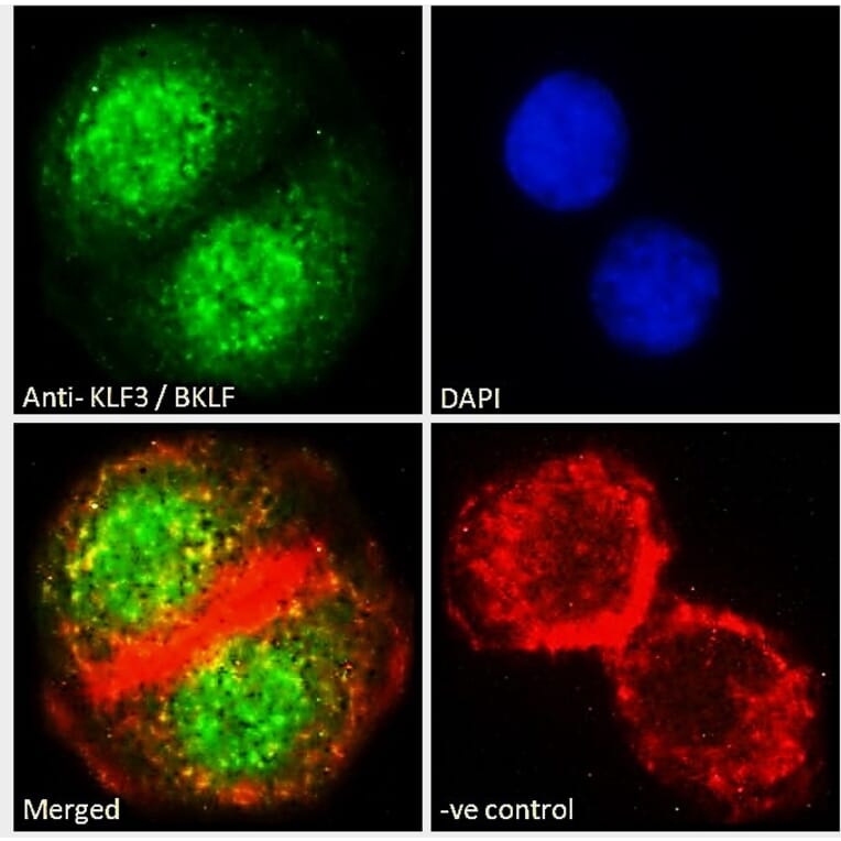
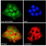
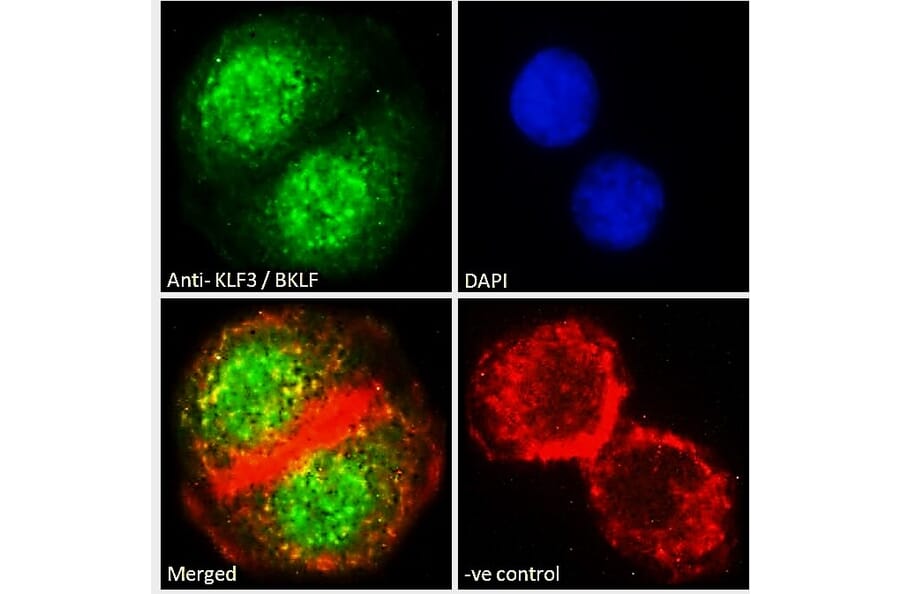
KLF3 expression in A549 cells analyzed by immunofluorescence. Cells were permeabilized with 0.15% Triton. Staining was performed with Anti-KLF3 Antibody (A83826) at 10µg/ml for 1 hour and Alexa Fluor 488 secondary antibody at 2µg/ml. Nuclear staining shown and nuclei were stained with DAPI (blue) while actin filaments were stained with phalloidin (red). Negative control: Goat IgG Isotype Control at 10µg/ml followed by Alexa Fluor 488 secondary antibody at 2µg/ml.
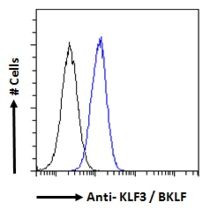
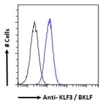
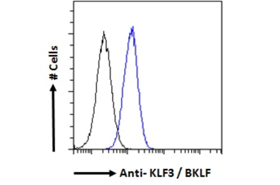
KLF3 expression in A431 cells (blue line) analyzed by flow cytometry. Cells were fixed in PFA and permeabilized with 0.5% Triton. Staining was performed with Anti-KLF3 Antibody (A83826) at 10µg/ml for 1 hour and Alexa Fluor 488 secondary antibody at 1µg/ml. Negative Control: Goat IgG Isotype Control (black line) followed by Alexa Fluor 488 secondary antibody.
Publishing research using Anti-KLF3 Antibody (A83826)? Please let us know so that we can list the citation on this page.
Alternative products to Anti-KLF3 Antibody (A83826)