Anti-Vitamin D Binding Protein Antibody [VDBP/4482]
Description
Mouse monoclonal [VDBP/4482] antibody to Vitamin D Binding Protein.
Specificity
Vitamin D-binding protein (DBP) is a multi-functional serum protein that binds to the plasma membranes of numerous cell types and mediates a variety of cellular functions. The locus of the DBP protein (also known as group-specific component protein or GC) is located at human chromosome 4q13.3. DBP functions in organ-specific transportation of vitamin D and its metabolites to the various target organs of the vitamin D endocrine system. In addition, DBP has immunomodulatory properties and is able to bind to the surface of leukocytes. DBP binds to the plasma membrane through a chondroitin sulfate proteoglycan. DBP serves as a co-chemotactic factor for C5a to enhance the chemotactic activity of C5a. DBP can also bind to globular Actin with high affinity and is involved in the clearance of Actin from the blood. DBP plays an important role in osteoclast differentiation. The diverse cellular functions of DBP require its cell surface binding ability to mediate different biological processes.
Applications
IHC-P
Dilutions
IHC-P: 1-2 µg/ml
Reactivity
Human
Immunogen
Recombinant fragment, around amino acids 35-175, of human Vitamin D Binding Protein. The exact sequence is proprietary.
Host
Mouse
Clonality
Monoclonal
Clone ID
VDBP/4482
Isotype
IgG2b
Light Chains
kappa
Conjugate
Unconjugated
Purification
Protein A/G chromatography.
Concentration
200 µg/ml
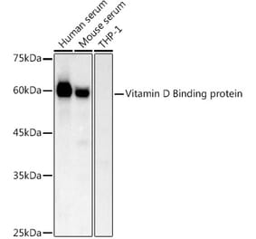
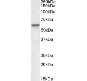
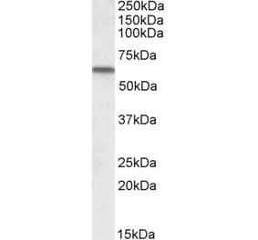
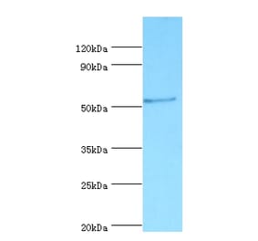
Molecular Weight
53 kDa
Product Form
Liquid
Formulation
Supplied in 10mM Phosphate Buffered Saline with 0.05% BSA and 0.05% Sodium Azide.
Storage
Shipped at 4°C. Upon delivery aliquot and store at -20°C. Avoid freeze / thaw cycles.
SDS-PAGE - Anti-Vitamin D Binding Protein Antibody [VDBP/4482] (A277626)
SDS-PAGE analysis of Anti-Vitamin D Binding Protein Antibody [VDBP/4482] under non-reduced and reduced conditions; showing intact IgG and intact heavy and light chains, respectively. SDS-PAGE analysis confirms the integrity and purity of the antibody.
Immunohistochemistry - Anti-Vitamin D Binding Protein Antibody [VDBP/4482] (A277626)
Immunohistochemical analysis of formalin-fixed, paraffin-embedded human tonsil using Anti-Vitamin D Binding Protein Antibody [VDBP/4482] at 2µg/ml in PBS for 30 minutes at room temperature.
Immunohistochemistry - Anti-Vitamin D Binding Protein Antibody [VDBP/4482] (A277626)
Immunohistochemical analysis of formalin-fixed, paraffin-embedded human pancreas using Anti-Vitamin D Binding Protein Antibody [VDBP/4482] at 2µg/ml in PBS for 30 minutes at room temperature.
Anti-Vitamin D Binding Protein Antibody [VDBP/4482] (A277626)
Analysis of protein array containing more than 19,000 full-length human proteins using Anti-Vitamin D Binding Protein Antibody [VDBP/4482]. Z-Score and S- Score: The Z-score represents the strength of a signal that a monoclonal antibody (MAb) (in combination with a fluorescently-tagged anti-IgG secondary antibody) produces when binding to a particular protein on the HuProtTM array. Z-scores are described in units of standard deviations (SD's) above the mean value of all signals generated on that array. If targets on HuProtTM are arranged in descending order of the Z-score, the S-score is the difference (also in units of SD's) between the Z-score. S-score therefore represents the relative target specificity of a MAb to its intended target; a MAb is considered to be specific to its intended target, if the MAb has an S-score of at least 2.5. For example, if a MAb binds to protein X with a Z-score of 43 and to protein Y with a Z-score of 14, then the S-score for the binding of that MAb to protein X is equal to 29.
Publishing research using Anti-Vitamin D Binding Protein Antibody [VDBP/4482] (A277626)? Please let us know so that we can list the citation on this page.
Alternative products to Anti-Vitamin D Binding Protein Antibody [VDBP/4482] (A277626)


![SDS-PAGE - Anti-Vitamin D Binding Protein Antibody [VDBP/4482] (A277626) - Antibodies.com](https://cdn.antibodies.com/image/catalog/277/A277626_1.jpg?profile=product_top)
![Immunohistochemistry - Anti-Vitamin D Binding Protein Antibody [VDBP/4482] (A277626) - Antibodies.com](https://cdn.antibodies.com/image/catalog/277/A277626_2.jpg?profile=product_top)
![Immunohistochemistry - Anti-Vitamin D Binding Protein Antibody [VDBP/4482] (A277626) - Antibodies.com](https://cdn.antibodies.com/image/catalog/277/A277626_3.jpg?profile=product_top)
![Protein Array - Anti-Vitamin D Binding Protein Antibody [VDBP/4482] (A277626) - Antibodies.com](https://cdn.antibodies.com/image/catalog/277/A277626_4.jpg?profile=product_top)
![SDS-PAGE - Anti-Vitamin D Binding Protein Antibody [VDBP/4482] (A277626) - Antibodies.com](https://cdn.antibodies.com/image/catalog/277/A277626_1.jpg?profile=product_top_thumb)
![Immunohistochemistry - Anti-Vitamin D Binding Protein Antibody [VDBP/4482] (A277626) - Antibodies.com](https://cdn.antibodies.com/image/catalog/277/A277626_2.jpg?profile=product_top_thumb)
![Immunohistochemistry - Anti-Vitamin D Binding Protein Antibody [VDBP/4482] (A277626) - Antibodies.com](https://cdn.antibodies.com/image/catalog/277/A277626_3.jpg?profile=product_top_thumb)
![Protein Array - Anti-Vitamin D Binding Protein Antibody [VDBP/4482] (A277626) - Antibodies.com](https://cdn.antibodies.com/image/catalog/277/A277626_4.jpg?profile=product_top_thumb)
![SDS-PAGE - Anti-Vitamin D Binding Protein Antibody [VDBP/4482] (A277626) - Antibodies.com](https://cdn.antibodies.com/image/catalog/277/A277626_1.jpg?profile=product_image)
![Immunohistochemistry - Anti-Vitamin D Binding Protein Antibody [VDBP/4482] (A277626) - Antibodies.com](https://cdn.antibodies.com/image/catalog/277/A277626_2.jpg?profile=product_image)
![Immunohistochemistry - Anti-Vitamin D Binding Protein Antibody [VDBP/4482] (A277626) - Antibodies.com](https://cdn.antibodies.com/image/catalog/277/A277626_3.jpg?profile=product_image)
![Protein Array - Anti-Vitamin D Binding Protein Antibody [VDBP/4482] (A277626) - Antibodies.com](https://cdn.antibodies.com/image/catalog/277/A277626_4.jpg?profile=product_image)
![Western Blot - Anti-Vitamin D Binding protein Antibody [ARC53914] (A305367) - Antibodies.com](https://cdn.antibodies.com/image/catalog/305/A305367_1.jpg?profile=product_alternative)
![SDS-PAGE - Anti-Vitamin D Binding Protein Antibody [VDBP/4482] - BSA and Azide free (A278214) - Antibodies.com](https://cdn.antibodies.com/image/catalog/278/A278214_1.jpg?profile=product_alternative)
![SDS-PAGE - Anti-Vitamin D Binding Protein Antibody [VDBP/4481] (A277625) - Antibodies.com](https://cdn.antibodies.com/image/catalog/277/A277625_1.jpg?profile=product_alternative)
![SDS-PAGE - Anti-Vitamin D Binding Protein Antibody [VDBP/4481] - BSA and Azide free (A278213) - Antibodies.com](https://cdn.antibodies.com/image/catalog/278/A278213_1.jpg?profile=product_alternative)