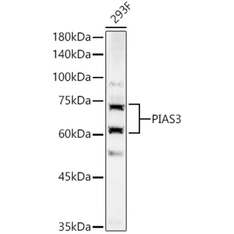
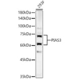
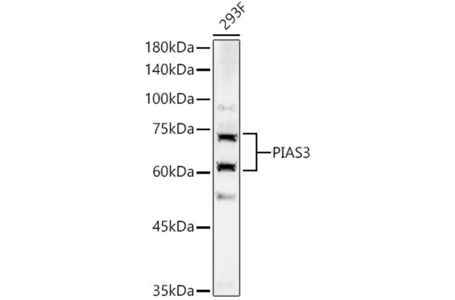
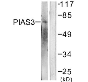
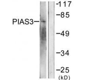
Western blot analysis of 293F, using Anti-PIAS3 Antibody (A16891) at 1:600 dilution. The secondary antibody was Goat Anti-Rabbit IgG H&L Antibody (HRP) at 1:10,000 dilution. Lysates/proteins were present at 25µg per lane. The blocking buffer used was 3% non-fat dry milk in TBST. Detection was with a ECL Basic Kit. Exposure time: 180s.
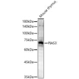
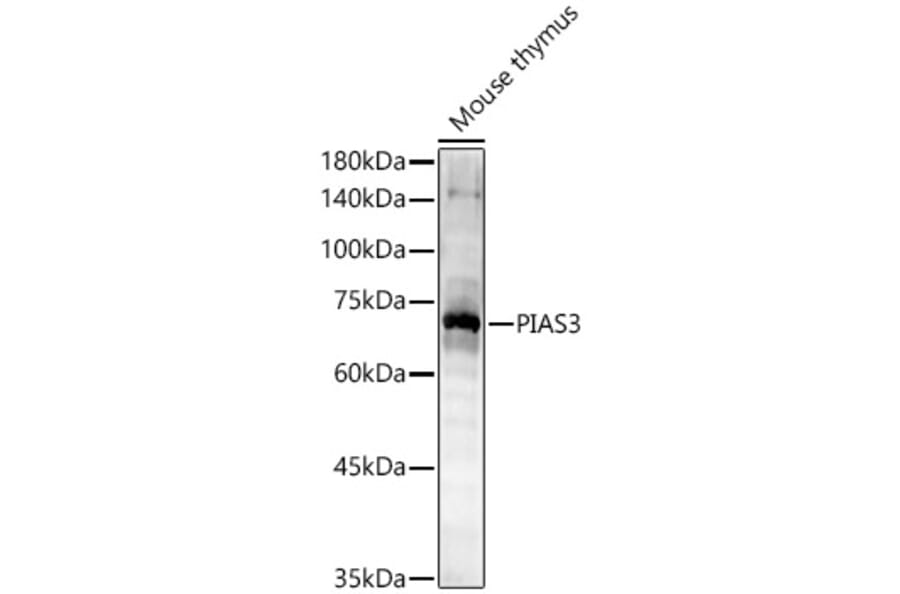
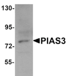
Western blot analysis of Mouse thymus, using Anti-PIAS3 Antibody (A16891) at 1:600 dilution. The secondary antibody was Goat Anti-Rabbit IgG H&L Antibody (HRP) at 1:10,000 dilution. Lysates/proteins were present at 25µg per lane. The blocking buffer used was 3% non-fat dry milk in TBST. Detection was with a ECL Basic Kit. Exposure time: 90s.
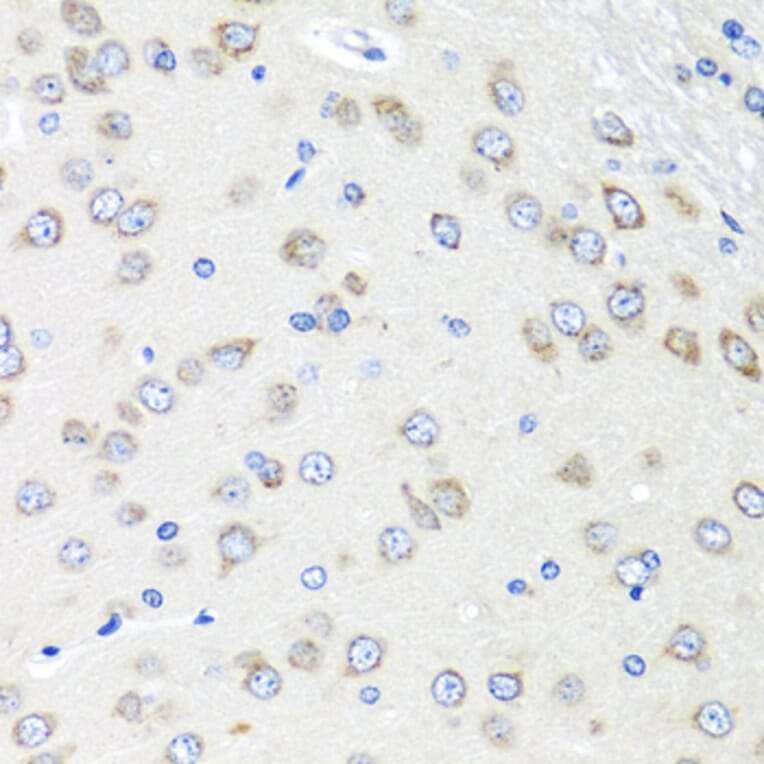
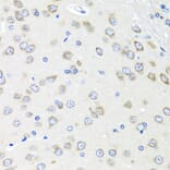
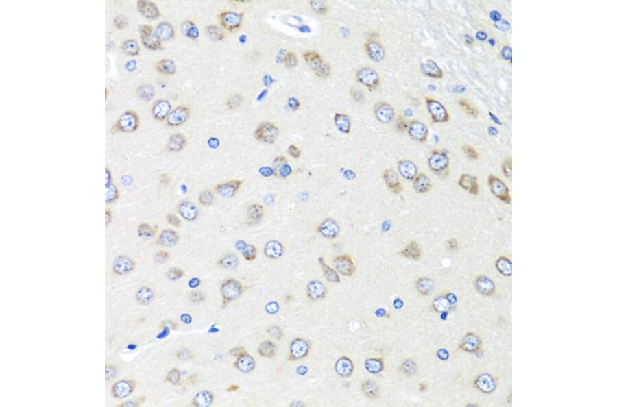
Immunohistochemistry analysis of paraffin-embedded rat brain using Anti-PIAS3 Antibody (A16891) at a dilution of 1:100 (40x lens). Perform microwave antigen retrieval with 10 mM PBS buffer pH 7.2 before commencing with IHC staining protocol.
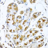
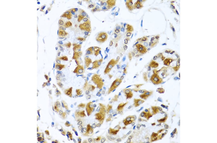
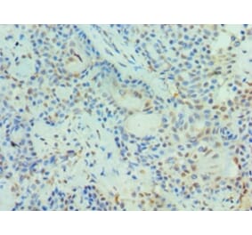
Immunohistochemistry analysis of paraffin-embedded human stomach using Anti-PIAS3 Antibody (A16891) at a dilution of 1:100 (40x lens). Perform microwave antigen retrieval with 10 mM PBS buffer pH 7.2 before commencing with IHC staining protocol.
Publishing research using Anti-PIAS3 Antibody (A16891)? Please let us know so that we can list the citation on this page.
Alternative products to Anti-PIAS3 Antibody (A16891)