Mouse monoclonal [G104] antibody to Phosphotyrosine.
Specificity
This antibody recognises phosphotyrosine, and detects the presence of phosphotyrosine in both un-stimulated and stimulated cell lysates.
Applications
WB, IHC, ICC/IF, IP
Dilutions
WB: 1:1,000, IHC: 1:100
Reactivity
Species Independent
Cross Reactivity
This antibody does not cross-react with phosphoserine or phosphothreonine.
Immunogen
Phosphotyrosine, alanine and glyceine in a 1:1:1 ratio polymerized in the presence of keyhole limpet hemocyanin with 1-ethyl-3-(3'-dimentrylaminopropyl) carbodiimide.
Host
Mouse
Clonality
Monoclonal
Clone ID
G104
Isotype
IgG1
Conjugate
Unconjugated
Purification
Protein G purification.
Concentration
1 mg/ml
Product Form
Liquid
Formulation
Supplied in Phosphate Buffered Saline, pH 7.4, with 50% Glycerol and 0.09% Sodium Azide.
Storage
Shipped at 4°C. Upon delivery aliquot and store at -20°C. Avoid freeze / thaw cycles.
Western Blot - Anti-Phosphotyrosine Antibody [G104] (A304807)
Western blot analysis of human A431 cell lysates showing detection of Phosphotyrosine protein using Anti-Phosphotyrosine Antibody [G104] (A304807) at 1:1,000 for 2 hours at room temperature. Load: 15 µg. Block: 1.5% BSA for 30 minutes at room temperature. The secondary antibody used was Sheep Anti-Mouse IgG: HRP for 1 hour at room temperature. Left: normal, right: EGF treated.
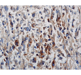
Immunohistochemistry analysis of mouse backskin, fixed in Bouin's fixative solution and paraffin-embedded. The Primary Antibody used was Anti-Phosphotyrosine Antibody [G104] (A304807) at 1:100 for 1 hour at room temperature. The secondary antibody used was FITC Goat Anti-Mouse (green) at 1:50 for 1 hour at room temperature. Localization: Stratum granulosum staining in the epidermis. Some dermal staining.
Publishing research using Anti-Phosphotyrosine Antibody [G104] (A304807)? Please let us know so that we can list the citation on this page.
Alternative products to Anti-Phosphotyrosine Antibody [G104] (A304807)


![Western Blot - Anti-Phosphotyrosine Antibody [G104] (A304807) - Antibodies.com](https://cdn.antibodies.com/image/catalog/304/A304807_1.png?profile=product_top)
![Immunohistochemistry - Anti-Phosphotyrosine Antibody [G104] (A304807) - Antibodies.com](https://cdn.antibodies.com/image/catalog/304/A304807_2.png?profile=product_top)
![Western Blot - Anti-Phosphotyrosine Antibody [G104] (A304807) - Antibodies.com](https://cdn.antibodies.com/image/catalog/304/A304807_1.png?profile=product_top_thumb)
![Immunohistochemistry - Anti-Phosphotyrosine Antibody [G104] (A304807) - Antibodies.com](https://cdn.antibodies.com/image/catalog/304/A304807_2.png?profile=product_top_thumb)
![Western Blot - Anti-Phosphotyrosine Antibody [G104] (A304807) - Antibodies.com](https://cdn.antibodies.com/image/catalog/304/A304807_1.png?profile=product_image)
![Immunohistochemistry - Anti-Phosphotyrosine Antibody [G104] (A304807) - Antibodies.com](https://cdn.antibodies.com/image/catalog/304/A304807_2.png?profile=product_image)
![Immunohistochemistry - Anti-Phosphotyrosine Antibody [PY20] - BSA and Azide free (A254036) - Antibodies.com](https://cdn.antibodies.com/image/catalog/254/A254038_1.jpg?profile=product_alternative)
![Immunohistochemistry - Anti-Phosphotyrosine Antibody [PY20] (A250856) - Antibodies.com](https://cdn.antibodies.com/image/catalog/250/A250858_1.jpg?profile=product_alternative)
![Immunohistochemistry - Anti-Phosphotyrosine Antibody [SPM102] (A250899) - Antibodies.com](https://cdn.antibodies.com/image/catalog/250/A250901_1.jpg?profile=product_alternative)
![Immunohistochemistry - Anti-Phosphotyrosine Antibody [SPM102] - BSA and Azide free (A254079) - Antibodies.com](https://cdn.antibodies.com/image/catalog/254/A254081_1.jpg?profile=product_alternative)
![SDS-PAGE - Anti-Phosphotyrosine Antibody [PY793] (A250935) - Antibodies.com](https://cdn.antibodies.com/image/catalog/250/A250935_1.jpg?profile=product_alternative)
![SDS-PAGE - Anti-Phosphotyrosine Antibody [PY793] - BSA and Azide free (A254115) - Antibodies.com](https://cdn.antibodies.com/image/catalog/254/A254115_1.jpg?profile=product_alternative)
![SDS-PAGE - Anti-Phosphotyrosine Antibody [PY265] - BSA and Azide free (A254134) - Antibodies.com](https://cdn.antibodies.com/image/catalog/254/A254135_1.jpg?profile=product_alternative)
![SDS-PAGE - Anti-Phosphotyrosine Antibody [PY265] (A250954) - Antibodies.com](https://cdn.antibodies.com/image/catalog/250/A250955_1.jpg?profile=product_alternative)
![SDS-PAGE - Anti-Phosphotyrosine Antibody [PY2870R] - BSA and Azide free (A254199) - Antibodies.com](https://cdn.antibodies.com/image/catalog/254/A254200_1.jpg?profile=product_alternative)
