

Unconjugated
Since the first report of induced pluripotent stem cells (iPSCs) by Takahashi and Yamanaka, numerous attempts have been made to derive iPSCs from other species via the ectopic expression of defined factors. Sheep iPSCs (siPSCs) have significant potential for biotechnology and agriculture. Although several groups have described siPSCs, the reprogramming efficiency was extremely low. The exogenous transgenes could be not silenced in the iPSCs, which hampered their development and application. Here, we report that p53 knockdown and antisilencing function 1A (ASF1A) overexpression promoted iPSC generation from sheep kidney cells (SKCs). Compared with transduction with eight human defined transcription factors (Oct4, Sox2, Klf4, c-Myc, Nanog, Lin28, hTERT, and SV40LT), the additional introduction of p53 RNA interference (RNAi) and/or ASF1A in the presence of small-molecule compounds [vitamin C (Vc) and valproic acid (VPA)] greatly improved the efficiency of sheep iPSC generation. The siPSCs exhibited morphological features similar to mouse embryonic stem cells (ESCs) and were positive for alkaline phosphatase and, pluripotent marker genes (Oct4, Nanog, Sox2, Rex1, TRA-1-60, TRA-1-81, and E-cadherin). Furthermore, these cells exhibited a normal karyotype of 54 chromosomes and were able to differentiate into all three germ layers both in vitro and in vivo. Moreover, the exogenous genes were silenced in siPSCs when p53 small hairpin RNA (shRNA) and ASF1A were added. Our results may help to reveal the role of p53 and ASF1A in sheep somatic cell reprogramming and provide an efficient approach to reprogramming sheep somatic cells.
We have investigated oxidized low-density lipoprotein (ox-LDL) induced senescence in hematopoietic stem cells (HCs). Mouse Sca-1+ HCs were separated and purified using the magnetic activated cell sorting technique. Ox-LDL induced significant senescence in HCs measured by SA-β-Gal staining, and reduced CFU-Mix colony-forming capacity, arresting cells at G0/G1 phase. In agreement with the cell cycle arrest, ox-LDL markedly reduced the expression of CDK4, cyclin D, and cyclin E. As possible contributing factors for cell senescence, ox-LDL also induced cellular oxidative stress and reduced telomerase activity.

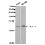
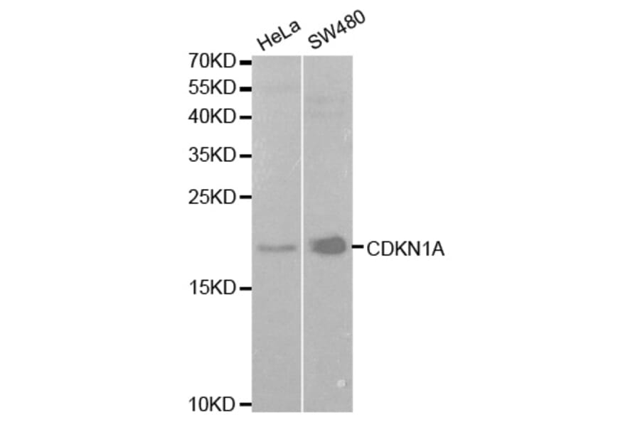


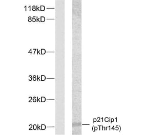
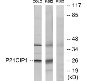
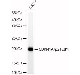
![Western Blot - Anti-p21 Antibody [ARC51040] (A307748) - Antibodies.com](https://cdn.antibodies.com/image/catalog/307/A307748_1.jpg?profile=product_alternative)
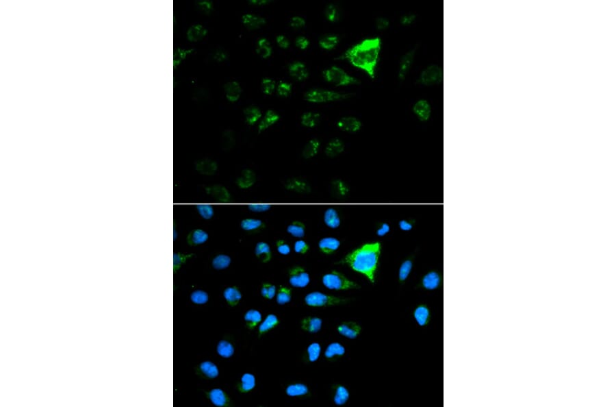
![Immunohistochemistry - Anti-p21 Antibody [WA-1] - BSA and Azide free (A251265) - Antibodies.com](https://cdn.antibodies.com/image/catalog/251/A251266_1.jpg?profile=product_alternative)
![Immunohistochemistry - Anti-p21 Antibody [DCS-60.2] - BSA and Azide free (A251266) - Antibodies.com](https://cdn.antibodies.com/image/catalog/251/A251267_1.jpg?profile=product_alternative)
![Immunohistochemistry - Anti-p21 Antibody [CIP1/823] (A248086) - Antibodies.com](https://cdn.antibodies.com/image/catalog/248/A248086_1.jpg?profile=product_alternative)
![Immunohistochemistry - Anti-p21 Antibody [HJ21] (A248086) - Antibodies.com](https://cdn.antibodies.com/image/catalog/248/A248087_1.jpg?profile=product_alternative)
![Immunohistochemistry - Anti-p21 Antibody [HJ21] - BSA and Azide free (A251269) - Antibodies.com](https://cdn.antibodies.com/image/catalog/251/A251270_1.jpg?profile=product_alternative)
![Immunohistochemistry - Anti-p21 Antibody [WA-1] (A248082) - Antibodies.com](https://cdn.antibodies.com/image/catalog/248/A248083_1.jpg?profile=product_alternative)
![Immunohistochemistry - Anti-p21 Antibody [DCS-60.2] (A248083) - Antibodies.com](https://cdn.antibodies.com/image/catalog/248/A248084_1.jpg?profile=product_alternative)