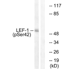
LEF1 expression in K562 nuclear cell lysate (A) + peptide (B) analyzed by western blot. Cells were lysed in RIPA buffer and 35µg protein was run per lane. Primary antibody incubation was performed with Anti-LEF1 Antibody (A84194) at 2µg/ml and detected by chemiluminescence.
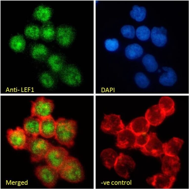
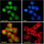
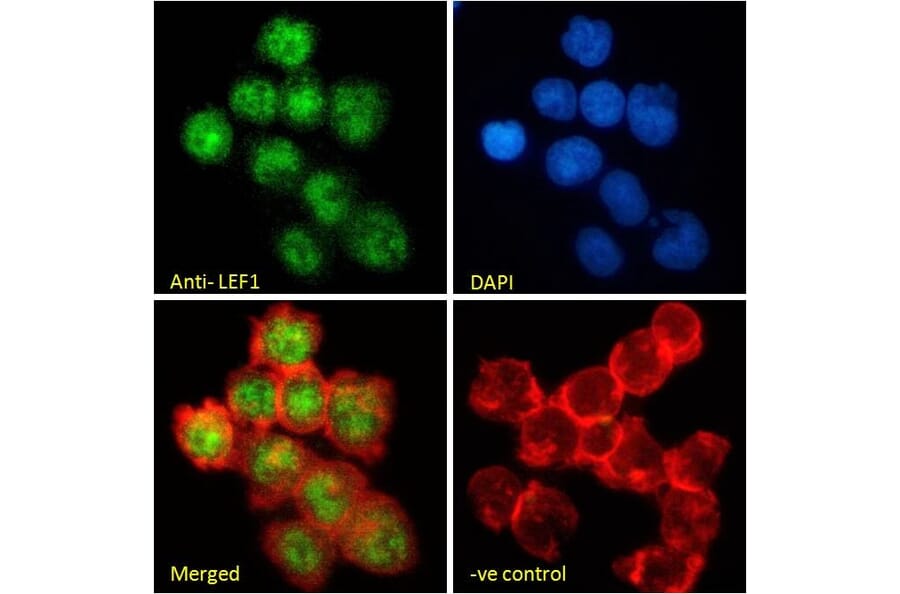
LEF1 expression in Jurkat cells analyzed by immunofluorescence. Cells were permeabilized with 0.15% Triton. Staining was performed with Anti-LEF1 Antibody (A84194) at 10µg/ml for 1 hour and Alexa Fluor 488 secondary antibody at 2µg/ml. Nuclear staining shown and nuclei were stained with DAPI (blue) while actin filaments were stained with phalloidin (red). Negative control: Goat IgG Isotype Control at 10µg/ml followed by Alexa Fluor 488 secondary antibody at 2µg/ml.
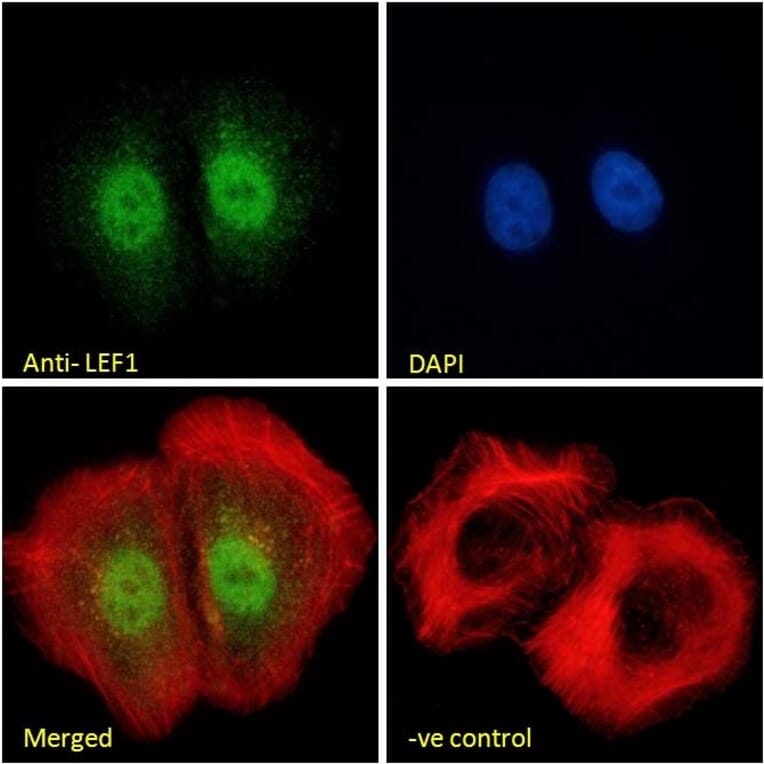
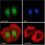
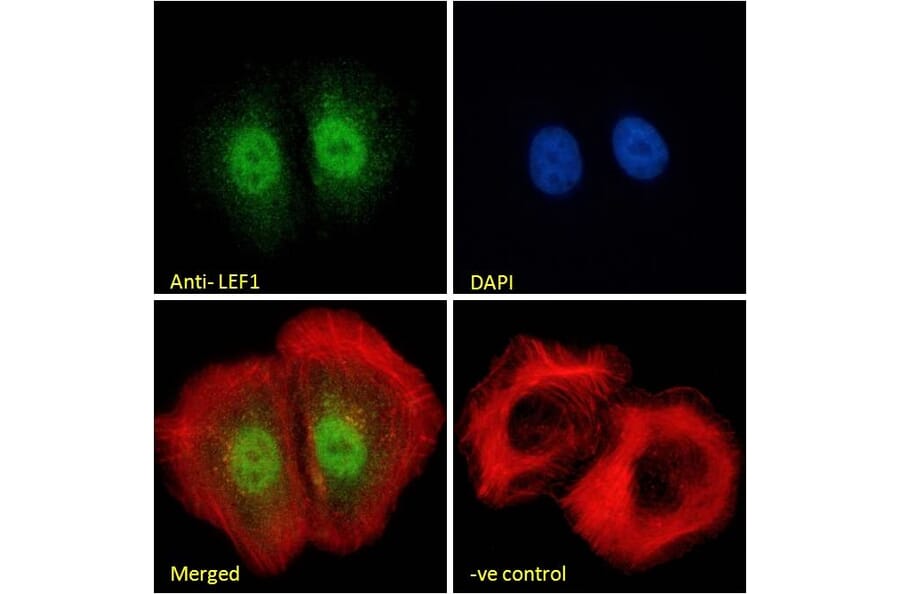
LEF1 expression in U2OS cells analyzed by immunofluorescence. Cells were permeabilized with 0.15% Triton. Staining was performed with Anti-LEF1 Antibody (A84194) at 10µg/ml for 1 hour and Alexa Fluor 488 secondary antibody at 2µg/ml. Nuclear staining shown and nuclei were stained with DAPI (blue) while actin filaments were stained with phalloidin (red). Negative control: Goat IgG Isotype Control at 10µg/ml followed by Alexa Fluor 488 secondary antibody at 2µg/ml.
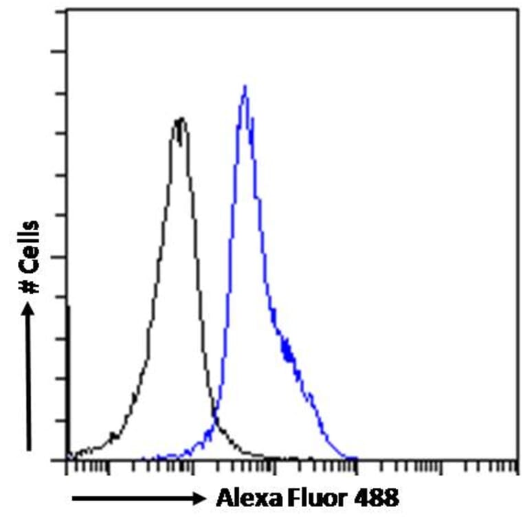
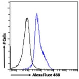
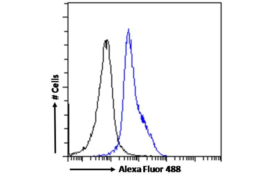
LEF1 expression in Jurkat cells (blue line) analyzed by flow cytometry. Cells were fixed in PFA and permeabilized with 0.5% Triton. Staining was performed with Anti-LEF1 Antibody (A84194) at 10µg/ml for 1 hour and Alexa Fluor 488 secondary antibody at 1µg/ml. Negative Control: Goat IgG Isotype Control (black line) followed by Alexa Fluor 488 secondary antibody.
Publishing research using Anti-LEF1 Antibody (A84194)? Please let us know so that we can list the citation on this page.
Alternative products to Anti-LEF1 Antibody (A84194)


![Western Blot - Anti-LEF1 Antibody [ARC1019] (A306507) - Antibodies.com](https://cdn.antibodies.com/image/catalog/306/A306507_1.jpg?profile=product_alternative)
![Immunohistochemistry - Anti-LEF1 Antibody [LEF1/341R] (A249581) - Antibodies.com](https://cdn.antibodies.com/image/catalog/249/A249582_1.jpg?profile=product_alternative)
![Immunohistochemistry - Anti-LEF1 Antibody [LEF1/341R] - BSA and Azide free (A252761) - Antibodies.com](https://cdn.antibodies.com/image/catalog/252/A252762_1.jpg?profile=product_alternative)
![Immunohistochemistry - Anti-LEF1 Antibody [rLEF1/6906] (A277907) - Antibodies.com](https://cdn.antibodies.com/image/catalog/277/A277907_1.jpg?profile=product_alternative)
![Immunohistochemistry - Anti-LEF1 Antibody [rLEF1/6906] - BSA and Azide free (A278495) - Antibodies.com](https://cdn.antibodies.com/image/catalog/278/A278495_1.jpg?profile=product_alternative)
![Immunohistochemistry - Anti-LEF1 Antibody [LEF1/422R] - BSA and Azide free (A252762) - Antibodies.com](https://cdn.antibodies.com/image/catalog/252/A252763_1.jpg?profile=product_alternative)
![Immunohistochemistry - Anti-LEF1 Antibody [LEF1/422R] (A249582) - Antibodies.com](https://cdn.antibodies.com/image/catalog/249/A249583_1.jpg?profile=product_alternative)
