Synthetic peptide from the N-terminal of Human IRGM.
Host
Rabbit
Clonality
Polyclonal
Isotype
IgG
Conjugate
Unconjugated
Purification
Peptide affinity purification.
Concentration
1 mg/ml
Molecular Weight
~20 kDa
Product Form
Liquid
Formulation
Supplied in Phosphate Buffered Saline with 50% Glycerol and 0.09% Sodium Azide.
Storage
Shipped at 4°C. Upon delivery aliquot and store at -20°C. Avoid freeze / thaw cycles.
Synonyms
IFI1, Immunity-related GTPase family M protein, Immunity-related GTPase family M protein 1, Interferon-inducible protein 1, IRGM1, LPS-stimulated RAW 264.7 macrophage protein 47 homolog, LRG-47, LRG47
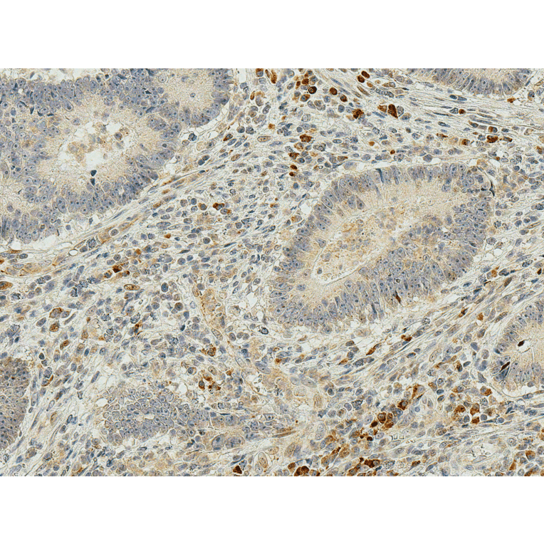
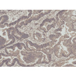
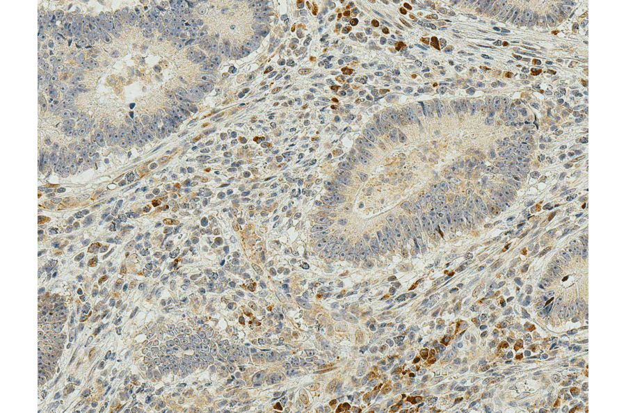
Immunohistochemistry analysis of human colon cancer, fixed in formalin and paraffin-embedded. The Primary Antibody used was Anti-IRGM Antibody (A304839) at 1:50 for 30 minutes at room temperature. Counterstain: Hematoxylin. Magnification: 20X. HRP-DAB Detection.
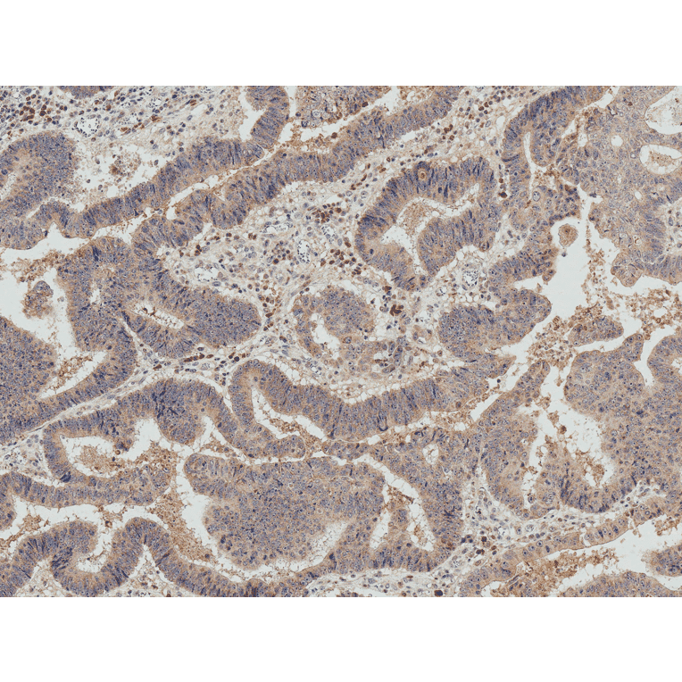
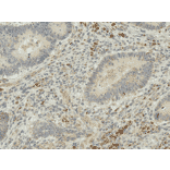
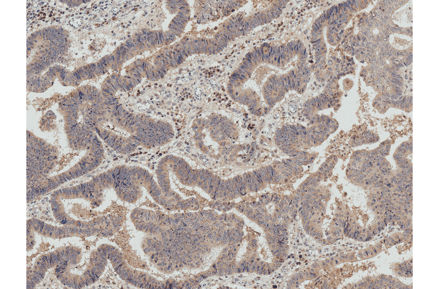
Immunohistochemistry analysis of human colon cancer, fixed in formalin and paraffin-embedded. The Primary Antibody used was Anti-IRGM Antibody (A304839) at 1:50 for 30 minutes at room temperature. Counterstain: Hematoxylin. Magnification: 10X. HRP-DAB Detection.
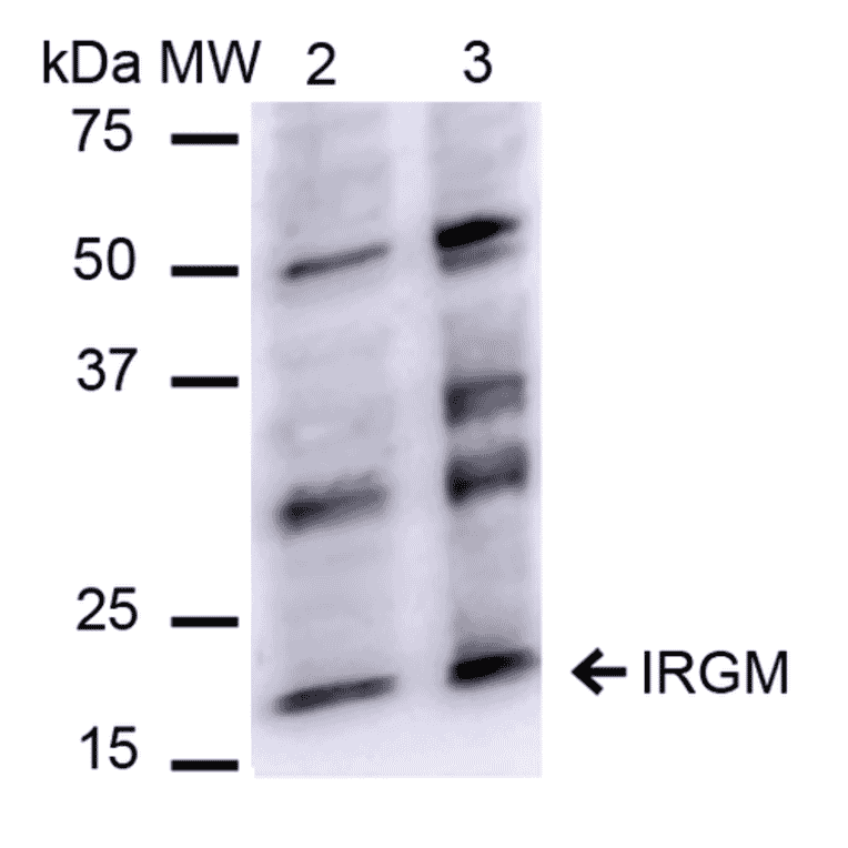
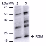
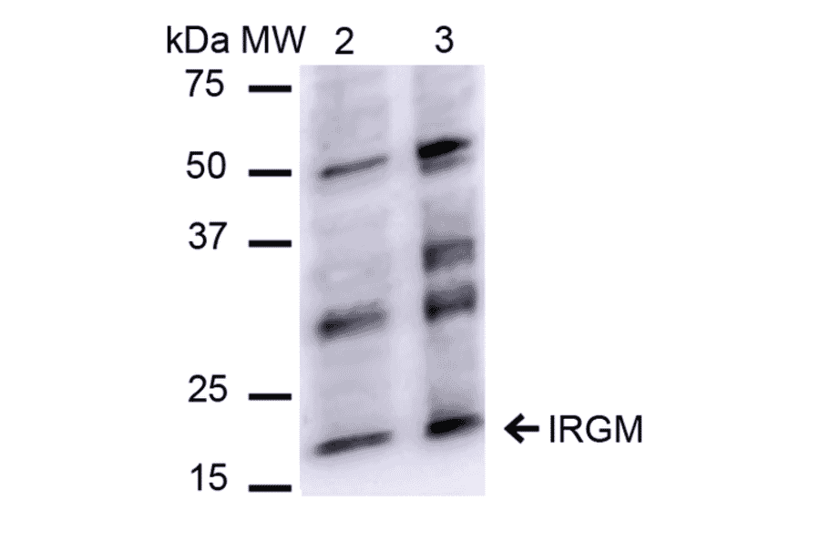
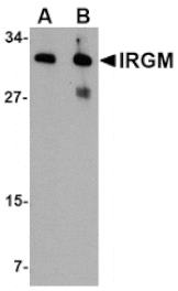
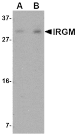
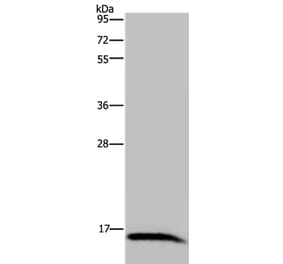
Western blot analysis of human HeLa and HEK293Trap cell lysates showing detection of ~20.1 kDa IRGM protein using Anti-IRGM Antibody (A304839) at 1:1,000 for 2 hours at room temperature. Lane 1: Molecular Weight Ladder (MW). Lane 2: HeLa cell lysates. Lane 3: 293Trap cell lysates. Load: 15 µg. Block: 5% Skim Milk in 1X TBST. The secondary antibody used was Goat Anti-Rabbit IgG: HRP at 1:1,000 for 60 minutes at room temperature. Color Development: ECL solution for 6 min in room temperature. Predicted/Observed Size: ~20.1 kDa.
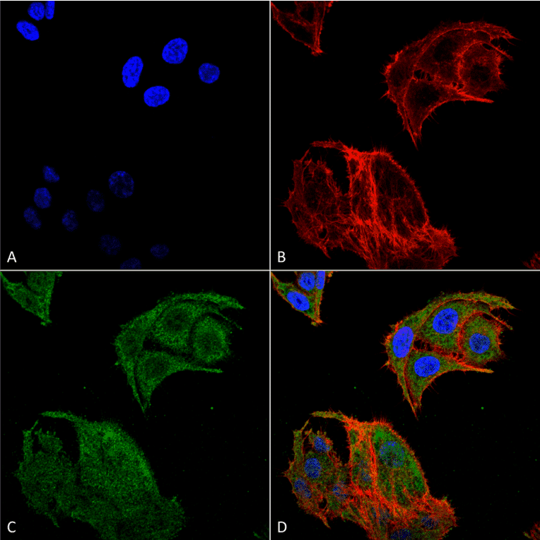
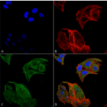
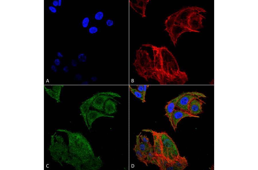
Immunocytochemistry/Immunofluorescence analysis of human colon carcinoma cell line (RKO), fixed in 4% formaldehyde for 15 min at room temperature, using Anti-IRGM Antibody (A304839), at 1:100 for 60 minutes at room temperature. The secondary antibody used was Goat Anti-Rabbit ATTO 488 at 1:100 for 60 minutes at room temperature. Counterstain: Phalloidin Texas Red F-Actin stain; DAPI (blue) nuclear stain at 1:1000, 1:5,000 for 60 minutes at room temperature, 5 minutes at room temperature. Localization: Cell Membrane, Cytoplasmic Vesicle, Phagosome Membrane, Autophagosome Membrane. Magnification: 60X.(A) DAPI nuclear stain. (B) Phalloidin Texas Red F-Actin stain. (C) IRGM Antibody. (D) Composite.
Publishing research using Anti-IRGM Antibody (A304839)? Please let us know so that we can list the citation on this page.
Alternative products to Anti-IRGM Antibody (A304839)