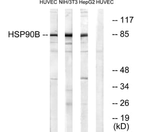
Western Blot - Anti-HSP90 beta Antibody [H9010] (A305130)
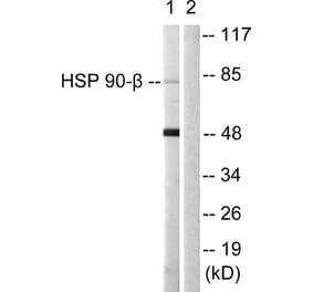
Western blot analysis of human Lysates showing detection of Hsp90 protein using Anti-HSP90 beta Antibody [H9010] (A305130) at 1:1,000. Comparison of clone H9010 behavior with Hsp90 human beta (1) and Hsp90 human alpha (2).
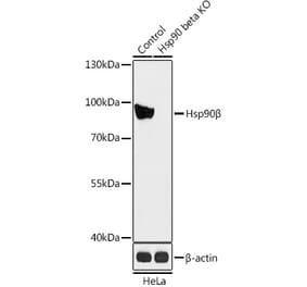
Western Blot - Anti-HSP90 beta Antibody [H9010] (A305130)
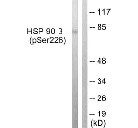
Western blot analysis of human Cervical cancer cell line (HeLa) lysate showing detection of Hsp90 protein using Anti-HSP90 beta Antibody [H9010] (A305130) at 1:1,000. The secondary antibody used was HRP Goat Anti-Mouse.
Immunohistochemistry analysis of mouse backskin, fixed in Bouin's fixative solution and paraffin-embedded. The Primary Antibody used was Anti-HSP90 beta Antibody [H9010] (A305130) at 1:100 for 1 hour at room temperature. The secondary antibody used was FITC Goat Anti-Mouse (green) at 1:50 for 1 hour at room temperature. Localization: Epidermis.
Immunohistochemistry analysis of mouse inflamed colon, fixed in formalin. The Primary Antibody used was Anti-HSP90 beta Antibody [H9010] (A305130) at 1:10,000 for 12 hours at 4°C. The secondary antibody used was Biotin Goat Anti-Mouse at 1:2000 for 1 hour at room temperature. Counterstain: Mayer Hematoxylin (purple/blue) nuclear stain at 200 µl for 2 minutes at room temperature. Localization: Inflammatory cells. Magnification: 40x. This image was produced using an amplifying IHC wash buffer. The antibody has therefore been diluted more than is recommended for other applications..
Immunohistochemistry analysis of human colon carcinoma, fixed in formalin. The Primary Antibody used was Anti-HSP90 beta Antibody [H9010] (A305130) at 1:10,000 for 12 hours at 4°C. The secondary antibody used was Alexa Fluor 555 Goat Anti-Mouse (red) at 1:5,000 for 1 hour at room temperature. Magnification: 40x. This image was produced using an amplifying IHC wash buffer. The antibody has therefore been diluted more than is recommended for other applications..
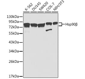
Western Blot - Anti-HSP90 beta Antibody [H9010] (A305130)
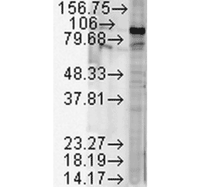
Western blot analysis of human cell lysates from various cell lines showing detection of Hsp90 protein using Anti-HSP90 beta Antibody [H9010] (A305130) at 1:1,000 for 2 hours at room temperature. Load: 15 µg. Block: 1.5% BSA for 30 minutes at room temperature. The secondary antibody used was Sheep Anti-Mouse IgG: HRP for 1 hour at room temperature.
Immunohistochemistry analysis of human colon carcinoma, fixed in formalin. The Primary Antibody used was Anti-HSP90 beta Antibody [H9010] (A305130) at 1:10,000 for 12 hours at 4°C. The secondary antibody used was Biotin Goat Anti-Mouse at 1:2000 for 1 hour at room temperature. Counterstain: Mayer Hematoxylin (purple/blue) nuclear stain at 200 µl for 2 minutes at room temperature. Localization: Inflammatory cells. Magnification: 40x. This image was produced using an amplifying IHC wash buffer. The antibody has therefore been diluted more than is recommended for other applications..
Immunohistochemistry analysis of mouse inflamed colon, fixed in formalin. The Primary Antibody used was Anti-HSP90 beta Antibody [H9010] (A305130) at 1:10,000 for 12 hours at 4°C. The secondary antibody used was Alexa Fluor 555 Goat Anti-Mouse (red) at 1:5,000 for 1 hour at room temperature. Localization: Inflammatory and epithelial mucosa. Magnification: 40x. Inflammatory and epithelial mucosa. This image was produced using an amplifying IHC wash buffer. The antibody has therefore been diluted more than is recommended for other applications..


![Western Blot - Anti-HSP90 beta Antibody [H9010] (A305130) - Antibodies.com](https://cdn.antibodies.com/image/catalog/305/A305130_1.png?profile=product_top)
![Western Blot - Anti-HSP90 beta Antibody [H9010] (A305130) - Antibodies.com](https://cdn.antibodies.com/image/catalog/305/A305130_2.png?profile=product_top)
![Immunohistochemistry - Anti-HSP90 beta Antibody [H9010] (A305130) - Antibodies.com](https://cdn.antibodies.com/image/catalog/305/A305130_3.png?profile=product_top)
![Immunohistochemistry - Anti-HSP90 beta Antibody [H9010] (A305130) - Antibodies.com](https://cdn.antibodies.com/image/catalog/305/A305130_4.png?profile=product_top)
![Immunohistochemistry - Anti-HSP90 beta Antibody [H9010] (A305130) - Antibodies.com](https://cdn.antibodies.com/image/catalog/305/A305130_5.png?profile=product_top)
![Western Blot - Anti-HSP90 beta Antibody [H9010] (A305130) - Antibodies.com](https://cdn.antibodies.com/image/catalog/305/A305130_6.png?profile=product_top)
![Immunohistochemistry - Anti-HSP90 beta Antibody [H9010] (A305130) - Antibodies.com](https://cdn.antibodies.com/image/catalog/305/A305130_7.png?profile=product_top)
![Immunohistochemistry - Anti-HSP90 beta Antibody [H9010] (A305130) - Antibodies.com](https://cdn.antibodies.com/image/catalog/305/A305130_8.png?profile=product_top)
![Western Blot - Anti-HSP90 beta Antibody [H9010] (A305130) - Antibodies.com](https://cdn.antibodies.com/image/catalog/305/A305130_1.png?profile=product_top_thumb)
![Western Blot - Anti-HSP90 beta Antibody [H9010] (A305130) - Antibodies.com](https://cdn.antibodies.com/image/catalog/305/A305130_2.png?profile=product_top_thumb)
![Immunohistochemistry - Anti-HSP90 beta Antibody [H9010] (A305130) - Antibodies.com](https://cdn.antibodies.com/image/catalog/305/A305130_3.png?profile=product_top_thumb)
![Immunohistochemistry - Anti-HSP90 beta Antibody [H9010] (A305130) - Antibodies.com](https://cdn.antibodies.com/image/catalog/305/A305130_4.png?profile=product_top_thumb)
![Immunohistochemistry - Anti-HSP90 beta Antibody [H9010] (A305130) - Antibodies.com](https://cdn.antibodies.com/image/catalog/305/A305130_5.png?profile=product_top_thumb)
![Western Blot - Anti-HSP90 beta Antibody [H9010] (A305130) - Antibodies.com](https://cdn.antibodies.com/image/catalog/305/A305130_6.png?profile=product_top_thumb)
![Immunohistochemistry - Anti-HSP90 beta Antibody [H9010] (A305130) - Antibodies.com](https://cdn.antibodies.com/image/catalog/305/A305130_7.png?profile=product_top_thumb)
![Western Blot - Anti-HSP90 beta Antibody [H9010] (A305130) - Antibodies.com](https://cdn.antibodies.com/image/catalog/305/A305130_1.png?profile=product_image)
![Western Blot - Anti-HSP90 beta Antibody [H9010] (A305130) - Antibodies.com](https://cdn.antibodies.com/image/catalog/305/A305130_2.png?profile=product_image)
![Immunohistochemistry - Anti-HSP90 beta Antibody [H9010] (A305130) - Antibodies.com](https://cdn.antibodies.com/image/catalog/305/A305130_3.png?profile=product_image)
![Immunohistochemistry - Anti-HSP90 beta Antibody [H9010] (A305130) - Antibodies.com](https://cdn.antibodies.com/image/catalog/305/A305130_4.png?profile=product_image)
![Immunohistochemistry - Anti-HSP90 beta Antibody [H9010] (A305130) - Antibodies.com](https://cdn.antibodies.com/image/catalog/305/A305130_5.png?profile=product_image)
![Western Blot - Anti-HSP90 beta Antibody [H9010] (A305130) - Antibodies.com](https://cdn.antibodies.com/image/catalog/305/A305130_6.png?profile=product_image)
![Immunohistochemistry - Anti-HSP90 beta Antibody [H9010] (A305130) - Antibodies.com](https://cdn.antibodies.com/image/catalog/305/A305130_7.png?profile=product_image)
![Immunohistochemistry - Anti-HSP90 beta Antibody [H9010] (A305130) - Antibodies.com](https://cdn.antibodies.com/image/catalog/305/A305130_8.png?profile=product_image)

![Western Blot - Anti-HSP90 beta Antibody [Hyb-K3701] (A304736) - Antibodies.com](https://cdn.antibodies.com/image/catalog/304/A304736_1.png?profile=product_alternative)