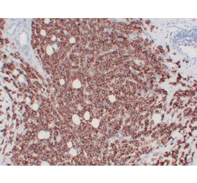
This antibody recognizes a protein of 185kDa, which is identified as c-erbB-2/HER-2/neu. Its epitope is localized in the extracellular domain. C-erbB-2/HER-2 is a member of the EGFR family. This MAb is specific and shows minimal cross-reaction with other members of the EGFR-family. Receptors of this family are located on the plasma membrane and consist of an extracellular ligand-binding domain that is connected to a large intracellular domain by a single transmembrane sequence. c-erbB-2/HER-2 protein is over-expressed in a variety of carcinomas especially those of breast and ovary.
SDS-PAGE analysis of Anti-HER2 Antibody [ERBB2/2452] under non-reduced and reduced conditions; showing intact IgG and intact heavy and light chains, respectively. SDS-PAGE analysis confirms the integrity and purity of the antibody.
Analysis of protein array containing more than 19,000 full-length human proteins using Anti-HER2 Antibody [ERBB2/2452]. Z-Score and S- Score: The Z-score represents the strength of a signal that a monoclonal antibody (MAb) (in combination with a fluorescently-tagged anti-IgG secondary antibody) produces when binding to a particular protein on the HuProtTM array. Z-scores are described in units of standard deviations (SD's) above the mean value of all signals generated on that array. If targets on HuProtTM are arranged in descending order of the Z-score, the S-score is the difference (also in units of SD's) between the Z-score. S-score therefore represents the relative target specificity of a MAb to its intended target; a MAb is considered to be specific to its intended target, if the MAb has an S-score of at least 2.5. For example, if a MAb binds to protein X with a Z-score of 43 and to protein Y with a Z-score of 14, then the S-score for the binding of that MAb to protein X is equal to 29.
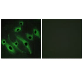
Immunofluorescent analysis of PFA fixed MCF-7 cells stained with Anti-HER2 Antibody [ERBB2/2452] followed by Goat Anti-Mouse IgG (CF® 488) (Green). Nuclear counterstain is RedDot.
Flow cytometric analysis of trypsinized PFA fixed MCF-7 cells using Anti-HER2 Antibody [ERBB2/2452] followed by Goat Anti-Mouse IgG (CF® 488) (Blue). Isotype Control (Red).


![Immunohistochemistry - Anti-HER2 Antibody [ERBB2/2452] (A248443) - Antibodies.com](https://cdn.antibodies.com/image/catalog/248/A248443_1.jpg?profile=product_top)
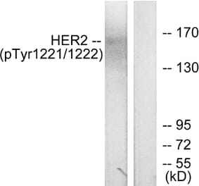
![SDS-PAGE - Anti-HER2 Antibody [ERBB2/2452] (A248443) - Antibodies.com](https://cdn.antibodies.com/image/catalog/248/A248443_2.jpg?profile=product_top)
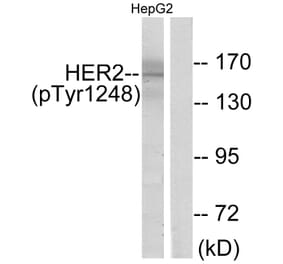
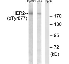
![Protein Array - Anti-HER2 Antibody [ERBB2/2452] (A248442) - Antibodies.com](https://cdn.antibodies.com/image/catalog/248/A248443_3.jpg?profile=product_top)
![Immunofluorescence - Anti-HER2 Antibody [ERBB2/2452] (A248443) - Antibodies.com](https://cdn.antibodies.com/image/catalog/248/A248443_4.jpg?profile=product_top)
![Flow Cytometry - Anti-HER2 Antibody [ERBB2/2452] (A248443) - Antibodies.com](https://cdn.antibodies.com/image/catalog/248/A248443_5.jpg?profile=product_top)
![Immunohistochemistry - Anti-HER2 Antibody [ERBB2/2452] (A248443) - Antibodies.com](https://cdn.antibodies.com/image/catalog/248/A248443_1.jpg?profile=product_top_thumb)
![SDS-PAGE - Anti-HER2 Antibody [ERBB2/2452] (A248443) - Antibodies.com](https://cdn.antibodies.com/image/catalog/248/A248443_2.jpg?profile=product_top_thumb)
![Protein Array - Anti-HER2 Antibody [ERBB2/2452] (A248442) - Antibodies.com](https://cdn.antibodies.com/image/catalog/248/A248443_3.jpg?profile=product_top_thumb)
![Immunofluorescence - Anti-HER2 Antibody [ERBB2/2452] (A248443) - Antibodies.com](https://cdn.antibodies.com/image/catalog/248/A248443_4.jpg?profile=product_top_thumb)
![Flow Cytometry - Anti-HER2 Antibody [ERBB2/2452] (A248443) - Antibodies.com](https://cdn.antibodies.com/image/catalog/248/A248443_5.jpg?profile=product_top_thumb)
![Immunohistochemistry - Anti-HER2 Antibody [ERBB2/2452] (A248443) - Antibodies.com](https://cdn.antibodies.com/image/catalog/248/A248443_1.jpg?profile=product_image)
![SDS-PAGE - Anti-HER2 Antibody [ERBB2/2452] (A248443) - Antibodies.com](https://cdn.antibodies.com/image/catalog/248/A248443_2.jpg?profile=product_image)
![Protein Array - Anti-HER2 Antibody [ERBB2/2452] (A248442) - Antibodies.com](https://cdn.antibodies.com/image/catalog/248/A248443_3.jpg?profile=product_image)
![Immunofluorescence - Anti-HER2 Antibody [ERBB2/2452] (A248443) - Antibodies.com](https://cdn.antibodies.com/image/catalog/248/A248443_4.jpg?profile=product_image)
![Flow Cytometry - Anti-HER2 Antibody [ERBB2/2452] (A248443) - Antibodies.com](https://cdn.antibodies.com/image/catalog/248/A248443_5.jpg?profile=product_image)
![Immunohistochemistry - Anti-HER2 Antibody [ERBB2/4376R] - BSA and Azide free (A278563) - Antibodies.com](https://cdn.antibodies.com/image/catalog/278/A278563_1.jpg?profile=product_alternative)
![Immunofluorescence - Anti-HER2 Antibody [HRB2/451] (A248448) - Antibodies.com](https://cdn.antibodies.com/image/catalog/248/A248448_1.jpg?profile=product_alternative)
![Immunofluorescence - Anti-HER2 Antibody [HRB2/451] - BSA and Azide free (A251630) - Antibodies.com](https://cdn.antibodies.com/image/catalog/251/A251630_1.jpg?profile=product_alternative)
![Immunohistochemistry - Anti-HER2 Antibody [ERBB2/4439] - BSA and Azide free (A251641) - Antibodies.com](https://cdn.antibodies.com/image/catalog/251/A251641_1.jpg?profile=product_alternative)
![Immunohistochemistry - Anti-HER2 Antibody [ERBB2/2452] - BSA and Azide free (A251625) - Antibodies.com](https://cdn.antibodies.com/image/catalog/251/A251625_1.jpg?profile=product_alternative)
![Flow Cytometry - Anti-HER2 Antibody [ERB2/776] (A248455) - Antibodies.com](https://cdn.antibodies.com/image/catalog/248/A248456_1.jpg?profile=product_alternative)
![Immunohistochemistry - Anti-HER2 Antibody [ZR5] (A248458) - Antibodies.com](https://cdn.antibodies.com/image/catalog/248/A248458_1.jpg?profile=product_alternative)