Fusion protein amino acids 375-770 (C-terminus) of rat GIT1.
Host
Mouse
Clonality
Monoclonal
Clone ID
S39B-8
Isotype
IgG1
Conjugate
Unconjugated
Purification
Protein G purification.
Concentration
1 mg/ml
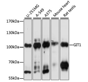
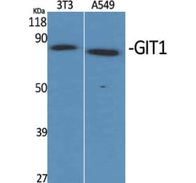
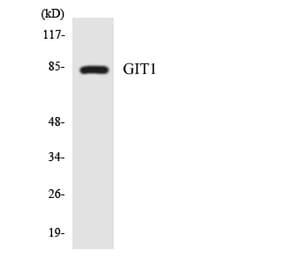
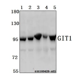
Molecular Weight
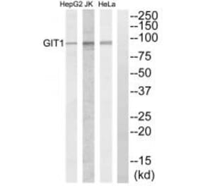
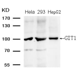
~90 kDa
Product Form
Liquid
Formulation
Supplied in Phosphate Buffered Saline, pH 7.4, with 50% Glycerol and 0.09% Sodium Azide.
Storage
Shipped at 4°C. Upon delivery aliquot and store at -20°C. Avoid freeze / thaw cycles.
Synonyms
ARF GAP GIT1, ARF GTPase-activating protein GIT1, CAT-1, CAT1, Cool-associated and tyrosine-phosphorylated protein 1, G protein-coupled receptor kinase-interactor 1, GRK-interacting protein 1, p95-APP1
Immunocytochemistry/Immunofluorescence analysis of human neuroblastoma cells (SH-SY5Y), fixed in 4% PFA for 15 min, using Anti-GIT1 Antibody [S39B-8] (A304764), at 1:50 for overnight at 4°C with slow rocking. The secondary antibody used was AlexaFluor 488 at 1:1,000 for 1 hour at room temperature. Counterstain: Phalloidin-iFluor 647 (red) F-Actin stain; Hoechst (blue) nuclear stain at 1:800, 1.6mM for 20 minutes at room temperature.(A) Hoechst (blue) nuclear stain. (B) Phalloidin-iFluor 647 (red) F-Actin stain. (C) GIT1 Antibody (D) Composite.
Immunocytochemistry/Immunofluorescence analysis of human neuroblastoma cell line (SK-N-BE, fixed in 4% formaldehyde for 15 min at room temperature, using Anti-GIT1 Antibody [S39B-8] (A304764), at 1:100 for 60 minutes at room temperature. The secondary antibody used was Goat Anti-Mouse ATTO 488 at 1:100 for 60 minutes at room temperature. Counterstain: Phalloidin Texas Red F-Actin stain; DAPI (blue) nuclear stain at 1:1000; 1:5,000 for 60 minutes room temperature, 5 minutes room temperature. Localization: Cytoplasm. Magnification: 60X.(A) DAPI (blue) nuclear stain. (B) Phalloidin Texas Red F-Actin stain. (C) GIT1 Antibody. (D) Composite.
Publishing research using Anti-GIT1 Antibody [S39B-8] (A304764)? Please let us know so that we can list the citation on this page.
Alternative products to Anti-GIT1 Antibody [S39B-8] (A304764)


![Immunocytochemistry/Immunofluorescence - Anti-GIT1 Antibody [S39B-8] (A304764) - Antibodies.com](https://cdn.antibodies.com/image/catalog/304/A304764_1.png?profile=product_top)
![Western Blot - Anti-GIT1 Antibody [S39B-8] (A304764) - Antibodies.com](https://cdn.antibodies.com/image/catalog/304/A304764_2.png?profile=product_top)
![Immunocytochemistry/Immunofluorescence - Anti-GIT1 Antibody [S39B-8] (A304764) - Antibodies.com](https://cdn.antibodies.com/image/catalog/304/A304764_3.png?profile=product_top)
![Immunocytochemistry/Immunofluorescence - Anti-GIT1 Antibody [S39B-8] (A304764) - Antibodies.com](https://cdn.antibodies.com/image/catalog/304/A304764_1.png?profile=product_top_thumb)
![Western Blot - Anti-GIT1 Antibody [S39B-8] (A304764) - Antibodies.com](https://cdn.antibodies.com/image/catalog/304/A304764_2.png?profile=product_top_thumb)
![Immunocytochemistry/Immunofluorescence - Anti-GIT1 Antibody [S39B-8] (A304764) - Antibodies.com](https://cdn.antibodies.com/image/catalog/304/A304764_3.png?profile=product_top_thumb)
![Immunocytochemistry/Immunofluorescence - Anti-GIT1 Antibody [S39B-8] (A304764) - Antibodies.com](https://cdn.antibodies.com/image/catalog/304/A304764_1.png?profile=product_image)
![Western Blot - Anti-GIT1 Antibody [S39B-8] (A304764) - Antibodies.com](https://cdn.antibodies.com/image/catalog/304/A304764_2.png?profile=product_image)
![Immunocytochemistry/Immunofluorescence - Anti-GIT1 Antibody [S39B-8] (A304764) - Antibodies.com](https://cdn.antibodies.com/image/catalog/304/A304764_3.png?profile=product_image)