Supplied in Phosphate Buffered Saline with 50% Glycerol and 5mM Sodium Azide.
Storage
Shipped at 4°C. Upon delivery aliquot and store at -20°C. Avoid freeze/thaw cycles.
General Notes
High quality antibodies to GFAP, like Anti-GFAP Antibody [5C10] (A85422), are useful for visualizing glia and monitoring developmental, disease, and damage related CNS alterations.
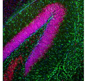
Immunofluorescent analysis of rat cerebellum section stained with Anti-GFAP Antibody [5C10] (A85422), dilution 1:1,000, in green, co-stained with Anti-NF-L Antibody (A85451), dilution 1:2,000, in red. Following transcardial perfusion with 4% paraformaldehyde, the brain was post-fixed for 24 hours, cut to 45 µm, and free-floating sections were stained with antibodies. Anti-GFAP Antibody [5C10] stains a network of astroglial cells, while the NF-L antibody labels neuronal cells and their processes.
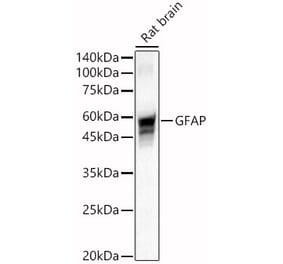
Western blot analysis of whole tissue lysates using Anti-GFAP Antibody [5C10] (A85422), dilution 1:2,000, in green. The lanes contain: [Lane 1] protein standard (red), [Lane 2] rat brain, [Lane 3] rat spinal cord, [Lane 4] mouse brain, [Lane 5] mouse spinal cord. The strong band at about 50 kDa corresponds to the GFAP protein.
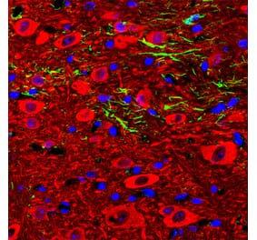
Mixed neuron-glial cultures stained with Anti-GFAP Antibody and Anti-NF-L Antibody (A85286 | green). The Anti-GFAP Antibody stains the network of astrocytes in these cultures, while the Anti-NF-L Antibody stains neurons and their processes. The blue channel shows the localization of DNA.
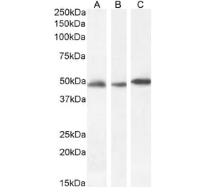
Strip blot of rat spinal cord protein extract stained with Anti-GFAP Antibody. A prominent band at about 50 kDa corresponds to the major isoform of GFAP.
Immunofluorescent analysis of cortical neuron-glial cell culture from E20 rat stained with Anti-Galectin 3 Antibody (A104321), at a dilution of 1:2,000, in green, and co-stained with Anti-GFAP Antibody [5C10] (A85422), at a dilution of 1:2,000, in red. The blue is Hoechst staining of nuclear DNA. Certain glial cells express Galectin 3 protein and so appear green, while the majority of glial cells and astrocytes produce GFAP protein and so appear red, and a few cells that express both proteins appear orange-yellow.
Immunohistochemistry analysis of paraffin embedded formalin fixed human cerebellum stained with Anti-GFAP Antibody [5C10] (A85422). The folds of the molecular layer are at the right and granular and white matter layers are on the left. Counterstained with Hematoxylin (blue).
Immunohistochemistry analysis of formalin fixed paraffin embedded adult horse brain samples removed whole, sliced incompletely cross ways and fixed over a 10-20 day period. The whole brain was put in a large necropsy specimen bucket containing 1-2 gallons of fixative, and after 24 hours, the formalin was poured off and then fresh formalin was added. The samples were fixed and embedded in paraffin in 2007 and sections were kept at room temperature until 2011. The sections were processed for antigen retrieval by boiling in Citrate buffer, pH 6, for 10 min. Primary incubation with Anti-GFAP Antibody [5C10] (A85422) was for 1 hour at 37°C, and secondary antibody incubation and color reaction was performed using the Vector mouse ABC kit.


![Immunofluorescence - Anti-GFAP Antibody [5C10] (A85422) - Antibodies.com](https://cdn.antibodies.com/image/catalog/85/A85422_1.jpg?profile=product_top)
![Western Blot - Anti-GFAP Antibody [5C10] (A85422) - Antibodies.com](https://cdn.antibodies.com/image/catalog/85/A85422_2.jpg?profile=product_top)
![Immunofluorescence - Anti-GFAP Antibody [5C10] (A85422) - Antibodies.com](https://cdn.antibodies.com/image/catalog/85/A85422_3.jpg?profile=product_top)
![Immunofluorescence - Anti-GFAP Antibody [5C10] (A85422) - Antibodies.com](https://cdn.antibodies.com/image/catalog/85/A85422_4.jpg?profile=product_top)
![Immunofluorescence - Anti-GFAP Antibody [5C10] (A85422) - Antibodies.com](https://cdn.antibodies.com/image/catalog/85/A85422_5.jpg?profile=product_top)
![Immunohistochemistry - Anti-GFAP Antibody [5C10] (A85422) - Antibodies.com](https://cdn.antibodies.com/image/catalog/85/A85422_6.jpg?profile=product_top)
![Immunohistochemistry - Anti-GFAP Antibody [5C10] (A85422) - Antibodies.com](https://cdn.antibodies.com/image/catalog/85/A85422_7.jpg?profile=product_top)
![Immunofluorescence - Anti-GFAP Antibody [5C10] (A85422) - Antibodies.com](https://cdn.antibodies.com/image/catalog/85/A85422_1.jpg?profile=product_top_thumb)
![Western Blot - Anti-GFAP Antibody [5C10] (A85422) - Antibodies.com](https://cdn.antibodies.com/image/catalog/85/A85422_2.jpg?profile=product_top_thumb)
![Immunofluorescence - Anti-GFAP Antibody [5C10] (A85422) - Antibodies.com](https://cdn.antibodies.com/image/catalog/85/A85422_3.jpg?profile=product_top_thumb)
![Immunofluorescence - Anti-GFAP Antibody [5C10] (A85422) - Antibodies.com](https://cdn.antibodies.com/image/catalog/85/A85422_4.jpg?profile=product_top_thumb)
![Immunofluorescence - Anti-GFAP Antibody [5C10] (A85422) - Antibodies.com](https://cdn.antibodies.com/image/catalog/85/A85422_5.jpg?profile=product_top_thumb)
![Immunohistochemistry - Anti-GFAP Antibody [5C10] (A85422) - Antibodies.com](https://cdn.antibodies.com/image/catalog/85/A85422_6.jpg?profile=product_top_thumb)
![Immunohistochemistry - Anti-GFAP Antibody [5C10] (A85422) - Antibodies.com](https://cdn.antibodies.com/image/catalog/85/A85422_7.jpg?profile=product_top_thumb)
![Immunofluorescence - Anti-GFAP Antibody [5C10] (A85422) - Antibodies.com](https://cdn.antibodies.com/image/catalog/85/A85422_1.jpg?profile=product_image)
![Western Blot - Anti-GFAP Antibody [5C10] (A85422) - Antibodies.com](https://cdn.antibodies.com/image/catalog/85/A85422_2.jpg?profile=product_image)
![Immunofluorescence - Anti-GFAP Antibody [5C10] (A85422) - Antibodies.com](https://cdn.antibodies.com/image/catalog/85/A85422_3.jpg?profile=product_image)
![Immunofluorescence - Anti-GFAP Antibody [5C10] (A85422) - Antibodies.com](https://cdn.antibodies.com/image/catalog/85/A85422_4.jpg?profile=product_image)
![Immunofluorescence - Anti-GFAP Antibody [5C10] (A85422) - Antibodies.com](https://cdn.antibodies.com/image/catalog/85/A85422_5.jpg?profile=product_image)
![Immunohistochemistry - Anti-GFAP Antibody [5C10] (A85422) - Antibodies.com](https://cdn.antibodies.com/image/catalog/85/A85422_6.jpg?profile=product_image)
![Immunohistochemistry - Anti-GFAP Antibody [5C10] (A85422) - Antibodies.com](https://cdn.antibodies.com/image/catalog/85/A85422_7.jpg?profile=product_image)
![Immunofluorescence - Anti-GFAP Antibody [2A5] (A104314) - Antibodies.com](https://cdn.antibodies.com/image/catalog/104/A104314_1.jpg?profile=product_alternative)
![Western Blot - Anti-GFAP Antibody [GA-5] - BSA and Azide free (A251887) - Antibodies.com](https://cdn.antibodies.com/image/catalog/251/A251887_1.jpg?profile=product_alternative)
![Immunohistochemistry - Anti-GFAP Antibody [GA-5 + ASTRO/789] (A248708) - Antibodies.com](https://cdn.antibodies.com/image/catalog/248/A248709_1.jpg?profile=product_alternative)
![Western Blot - Anti-GFAP Antibody [GA-5] (A248705) - Antibodies.com](https://cdn.antibodies.com/image/catalog/248/A248705_1.jpg?profile=product_alternative)
![Immunohistochemistry - Anti-GFAP Antibody [SPM248] - BSA and Azide free (A251888) - Antibodies.com](https://cdn.antibodies.com/image/catalog/251/A251888_1.jpg?profile=product_alternative)
![Immunohistochemistry - Anti-GFAP Antibody [SPM507] - BSA and Azide free (A251888) - Antibodies.com](https://cdn.antibodies.com/image/catalog/251/A251889_1.jpg?profile=product_alternative)
![Immunohistochemistry - Anti-GFAP Antibody [GA-5 + ASTRO/789] - BSA and Azide free (A251890) - Antibodies.com](https://cdn.antibodies.com/image/catalog/251/A251891_1.jpg?profile=product_alternative)
![Western Blot - Anti-GFAP Antibody [ARC0206] (A307282) - Antibodies.com](https://cdn.antibodies.com/image/catalog/307/A307282_1.jpg?profile=product_alternative)
![Immunohistochemistry - Anti-GFAP Antibody [SPM248] (A248706) - Antibodies.com](https://cdn.antibodies.com/image/catalog/248/A248706_1.jpg?profile=product_alternative)
![Immunohistochemistry - Anti-GFAP Antibody [ASTRO/789] (A248708) - Antibodies.com](https://cdn.antibodies.com/image/catalog/248/A248708_1.jpg?profile=product_alternative)
![Immunohistochemistry - Anti-GFAP Antibody [ASTRO/789] - BSA and Azide free (A251890) - Antibodies.com](https://cdn.antibodies.com/image/catalog/251/A251890_1.jpg?profile=product_alternative)
