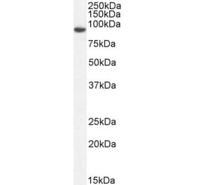
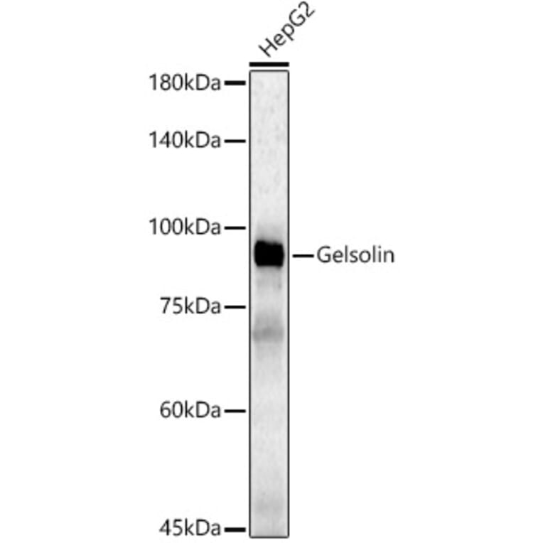
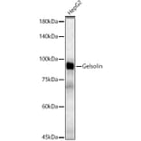
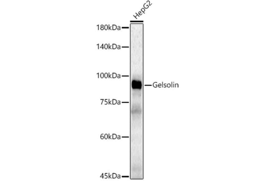
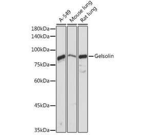
Western blot analysis of HepG2, using Anti-Gelsolin Antibody (A305452) at 1:500 dilution. The secondary antibody was Goat Anti-Rabbit IgG H&L Antibody (HRP) at 1:10,000 dilution. Lysates/proteins were present at 25µg per lane. The blocking buffer used was 3% non-fat dry milk in TBST. Detection was with a ECL Basic Kit. Exposure time: 90s.
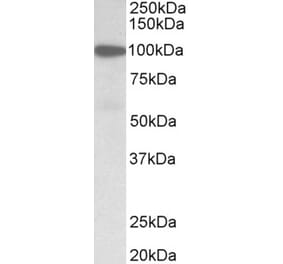
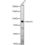
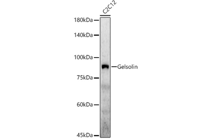
Western blot analysis of C2C12, using Anti-Gelsolin Antibody (A305452) at 1:500 dilution. The secondary antibody was Goat Anti-Rabbit IgG H&L Antibody (HRP) at 1:10,000 dilution. Lysates/proteins were present at 25µg per lane. The blocking buffer used was 3% non-fat dry milk in TBST. Detection was with a ECL Enhanced Kit (RM00021). Exposure time: 60s.
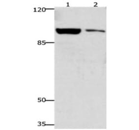
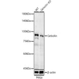
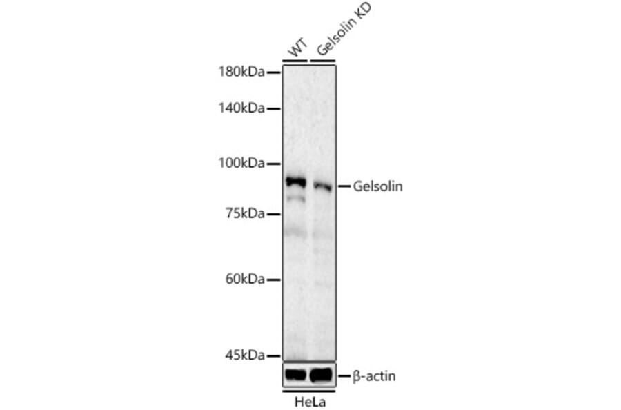
Western blot analysis of extracts from wild type(WT) and Gelsolin Rabbit polyclonal antibody knockdown (KD) HeLa cells, using Anti-Gelsolin Antibody (A305452) at 1:500 dilution. The secondary antibody was Goat Anti-Rabbit IgG H&L Antibody (HRP) at 1:10,000 dilution. Lysates/proteins were present at 25µg per lane. The blocking buffer used was 3% non-fat dry milk in TBST. Detection was with a ECL Basic Kit. Exposure time: 90s.
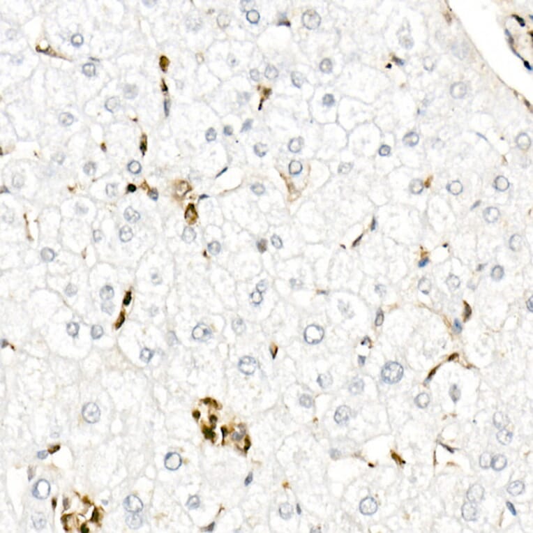
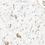
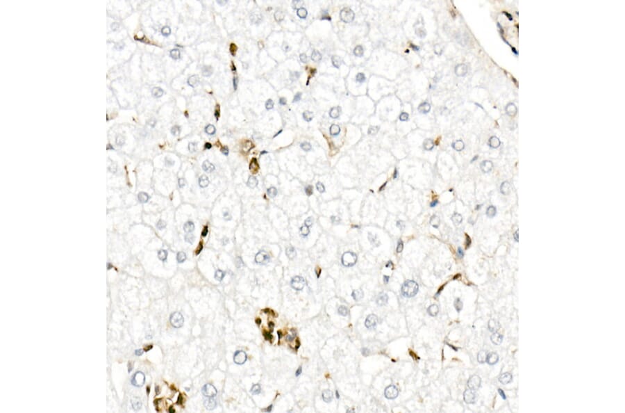
Immunohistochemistry analysis of paraffin-embedded human liver using Anti-Gelsolin Antibody (A305452) at a dilution of 1:50 (40x lens). Perform high pressure antigen retrieval with 10 mM citrate buffer pH 6.0 before commencing with IHC staining protocol.
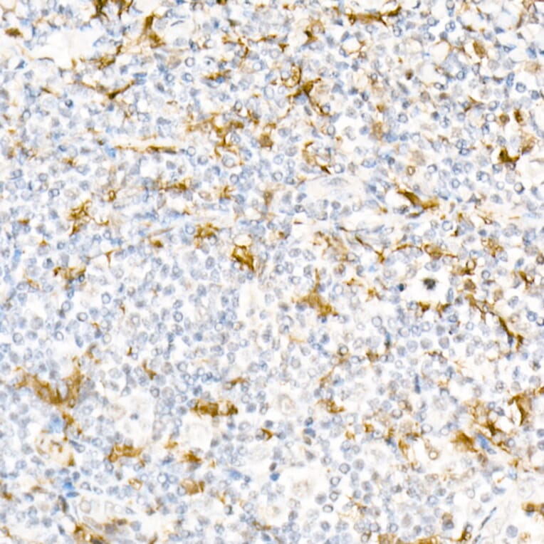
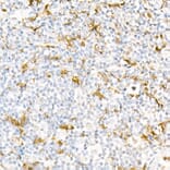
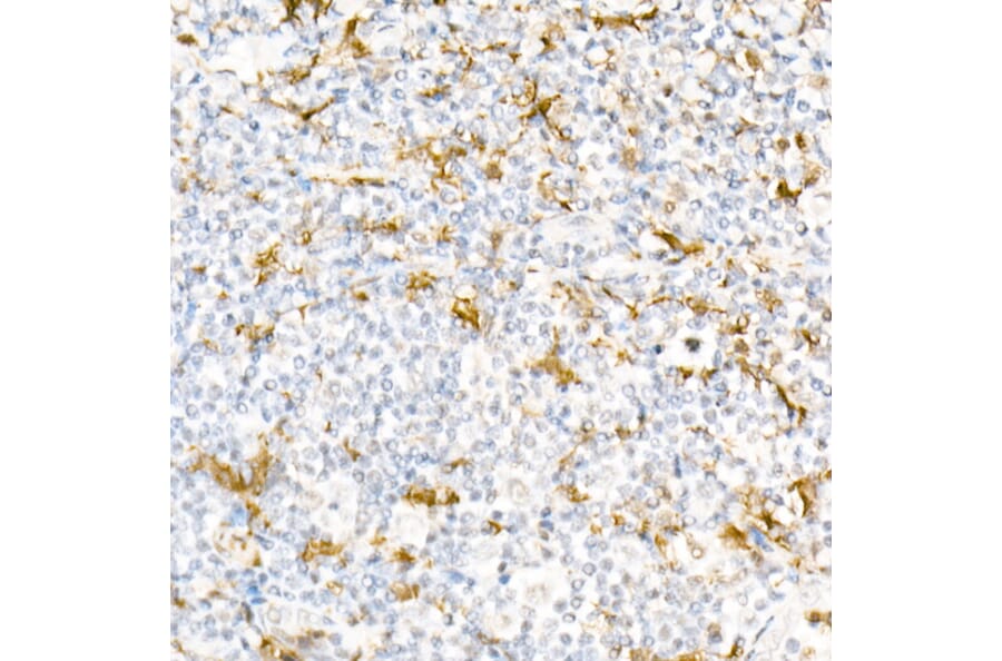
Immunohistochemistry analysis of paraffin-embedded human tonsil using Anti-Gelsolin Antibody (A305452) at a dilution of 1:50 (40x lens). Perform high pressure antigen retrieval with 10 mM citrate buffer pH 6.0 before commencing with IHC staining protocol.


![Western Blot - Anti-Gelsolin Antibody [ARC1924] (A305630) - Antibodies.com](https://cdn.antibodies.com/image/catalog/305/A305630_1.jpg?profile=product_alternative)