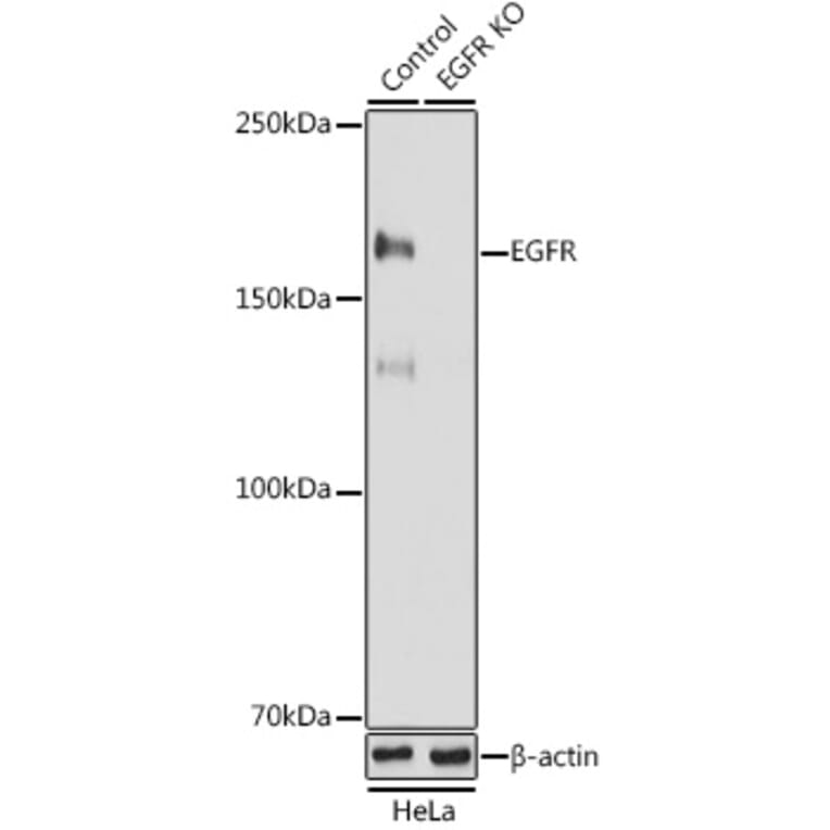
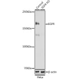
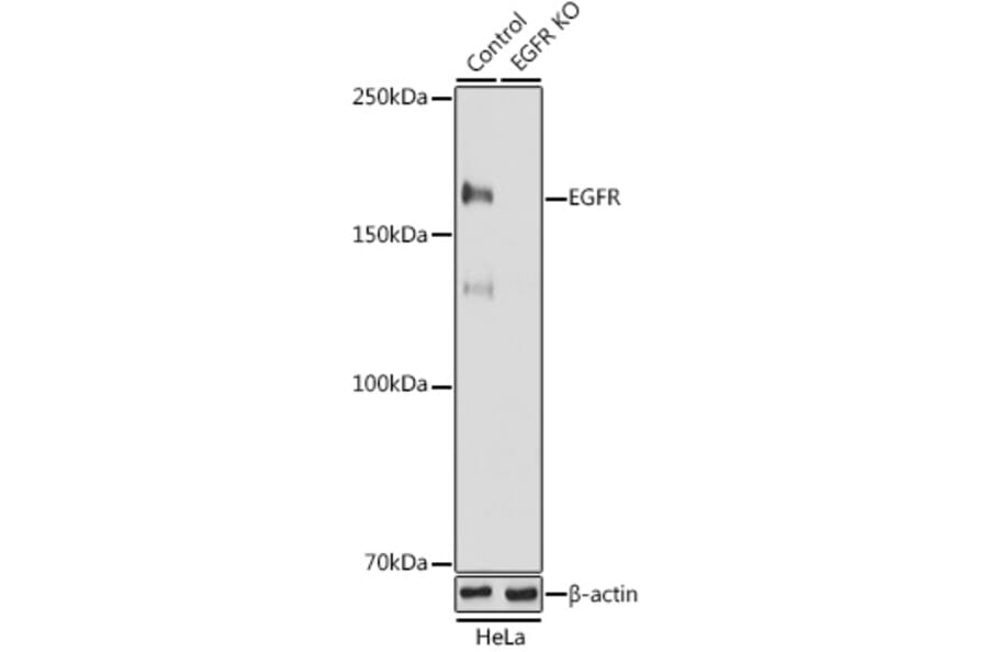
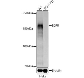
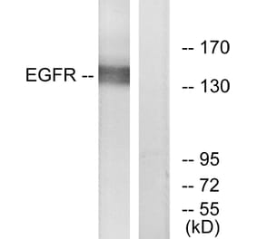
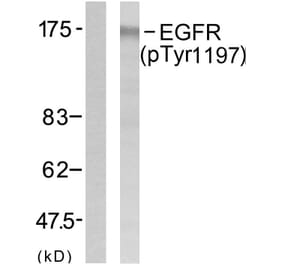
Western blot analysis of extracts from normal (control) and EGFR knockout (KO) HeLa cells, using Anti-EGFR Antibody (A81016) at 1:1,000 dilution. The secondary antibody was Goat Anti-Rabbit IgG H&L Antibody (HRP) at 1:10,000 dilution. Lysates/proteins were present at 25µg per lane. The blocking buffer used was 3% non-fat dry milk in TBST. Detection was with a ECL Basic Kit. Exposure time: 90s.
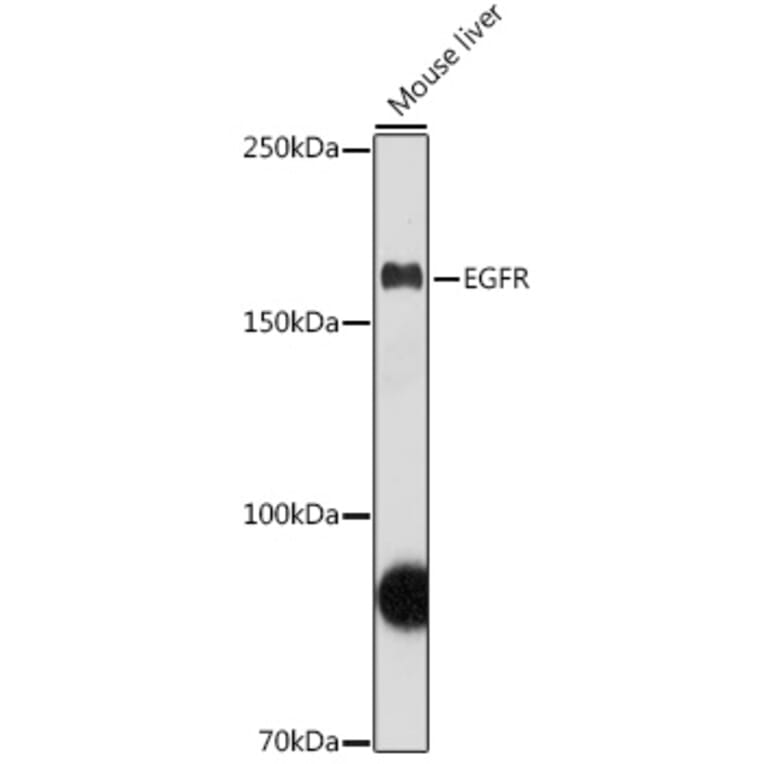
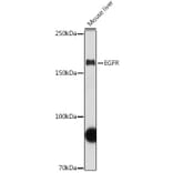
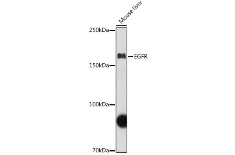
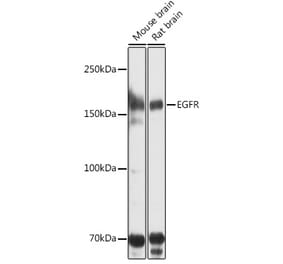
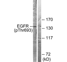
Western blot analysis of extracts of Mouse liver, using Anti-EGFR Antibody (A81016) at 1:1,000 dilution. The secondary antibody was Goat Anti-Rabbit IgG H&L Antibody (HRP) at 1:10,000 dilution. Lysates/proteins were present at 25µg per lane. The blocking buffer used was 3% non-fat dry milk in TBST. Detection was with a ECL Basic Kit. Exposure time: 90s.
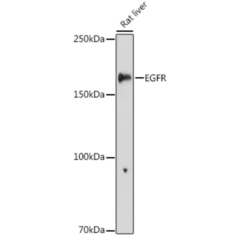
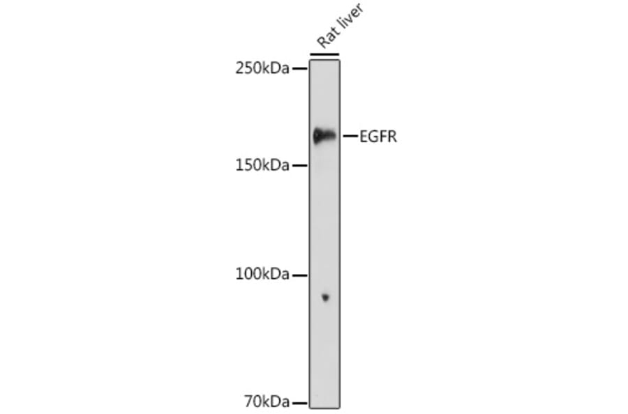
Western blot analysis of extracts of Rat liver, using Anti-EGFR Antibody (A81016) at 1:1,000 dilution. The secondary antibody was Goat Anti-Rabbit IgG H&L Antibody (HRP) at 1:10,000 dilution. Lysates/proteins were present at 25µg per lane. The blocking buffer used was 3% non-fat dry milk in TBST. Detection was with a ECL Enhanced Kit (RM00021). Exposure time: 180s.
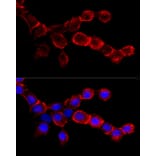
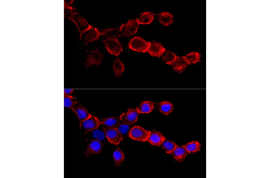
Immunofluorescence analysis of A-431 cells using Anti-EGFR Antibody (A81016) at a dilution of 1:50 (40x lens). DAPI was used to stain the cell nuclei (blue).
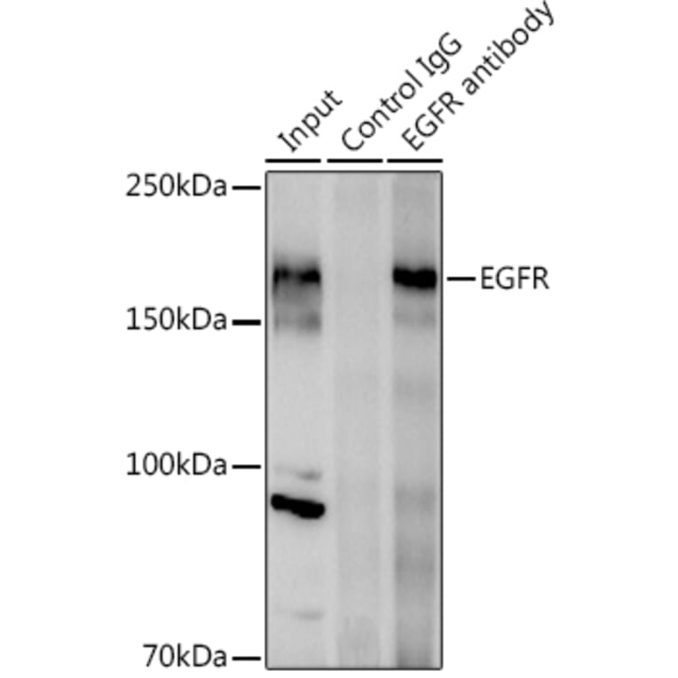
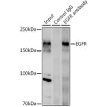
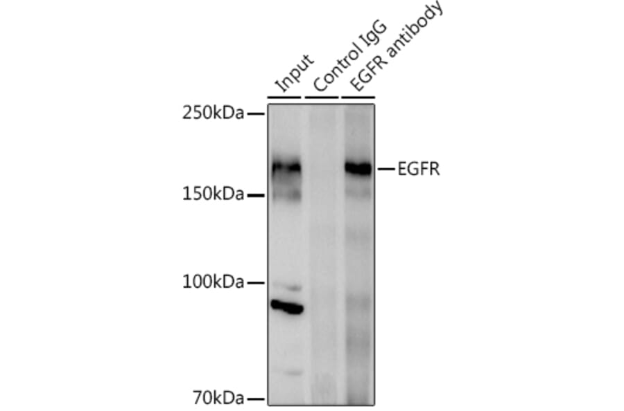
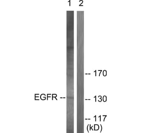
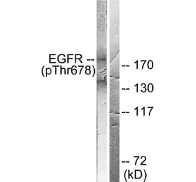
Immunoprecipitation analysis of 600µg extracts of mouse liver using 3µg of Anti-EGFR Antibody (A81016). This Western blot was performed on the immunoprecipitate using Anti-EGFR Antibody (A81016) at a dilution of 1:1000.