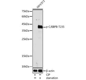
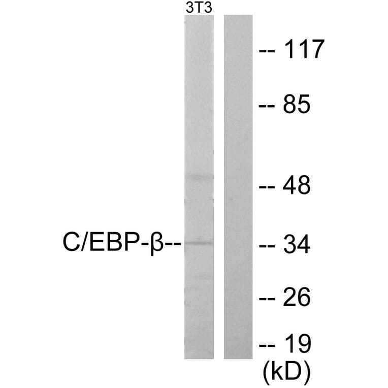
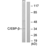
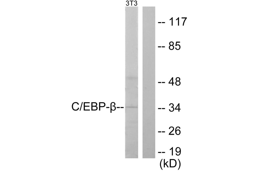
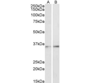
Western blot analysis of lysates from NIH/3T3 cells using Anti-CEBP beta Antibody. The right hand lane represents a negative control, where the antibody is blocked by the immunising peptide.
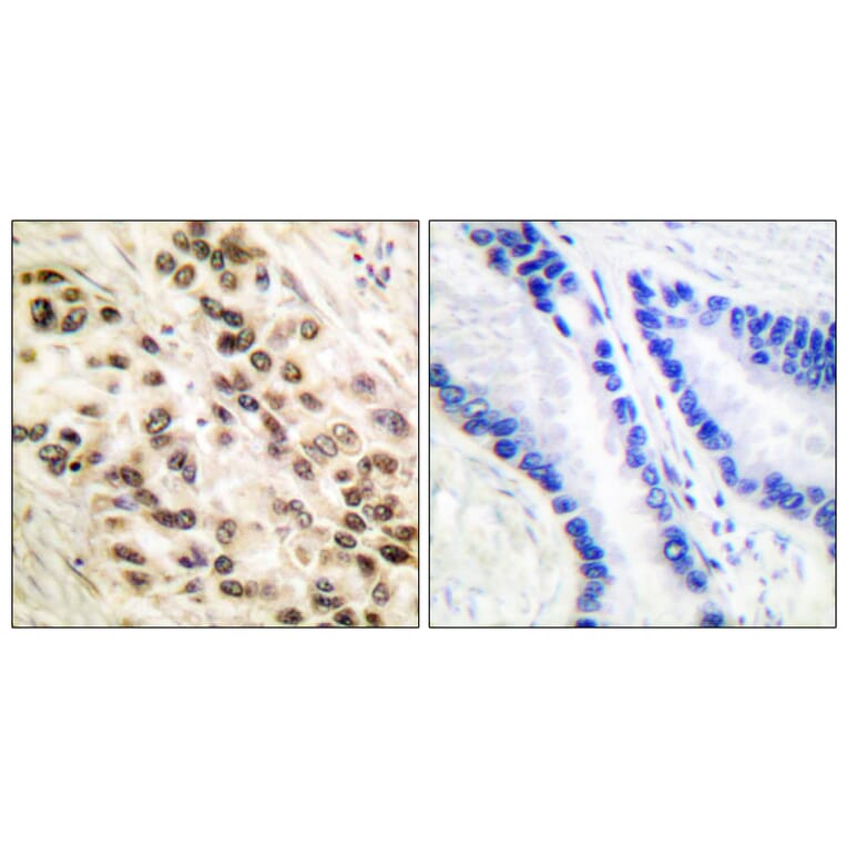
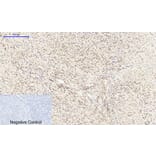
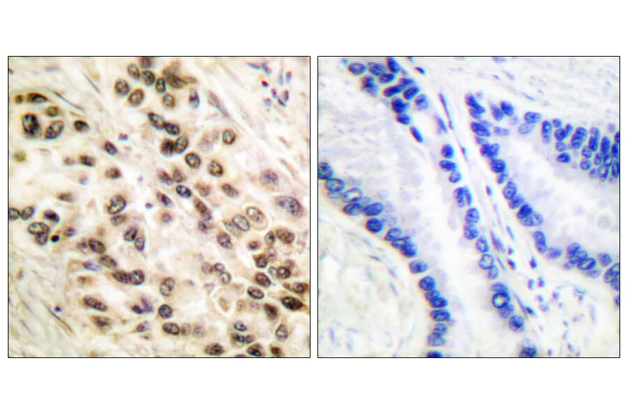
Immunohistochemical analysis of paraffin-embedded human lung carcinoma tissue using Anti-CEBP beta Antibody. The right hand panel represents a negative control, where the antibody was pre-incubated with the immunising peptide.
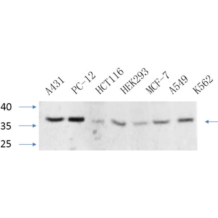
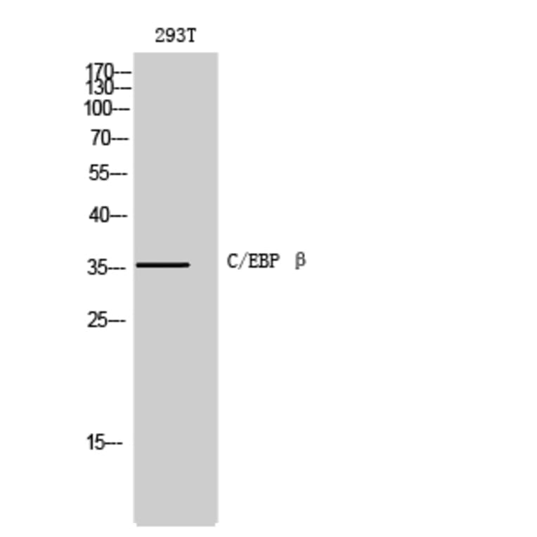
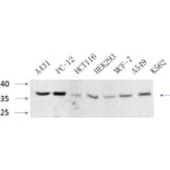
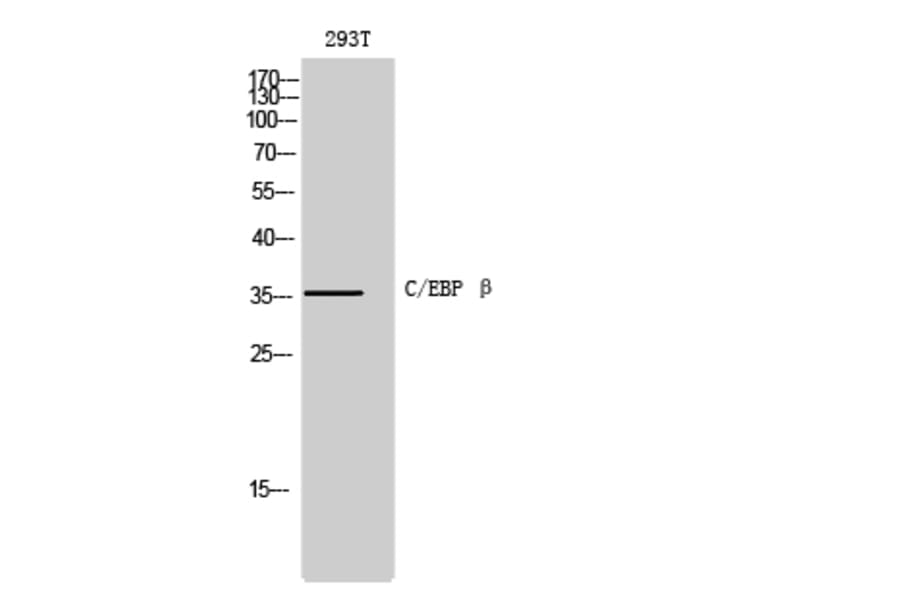
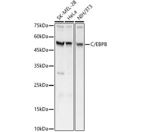
Western blot analysis of various cells using Anti-CEBP beta Antibody at 1:1,000 (4°C overnight). Goat Anti-Rabbit IgG (IRDye 800) was used as a secondary at 1:5,000 (25°C, 1 hour).
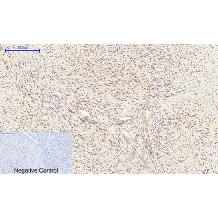
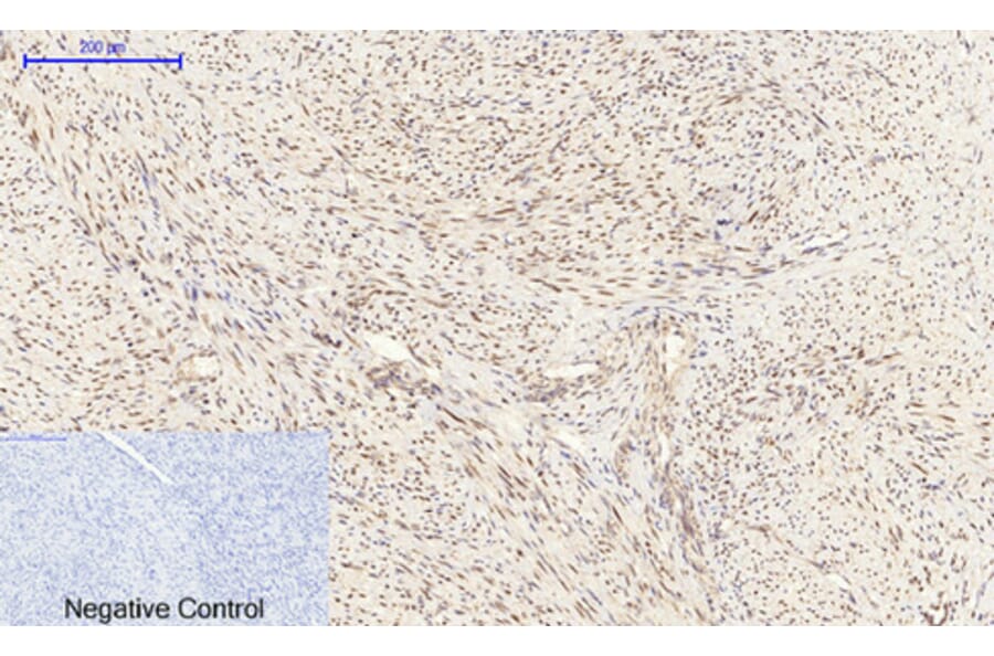
Immunohistochemical analysis of paraffin-embedded human uterus cancer tissue using Anti-CEBP beta Antibody at 1:200 (4°C overnight). Negative control was secondary antibody only.
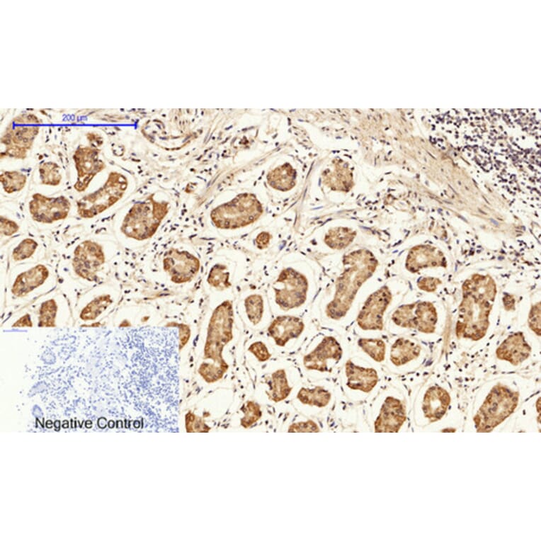
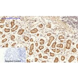
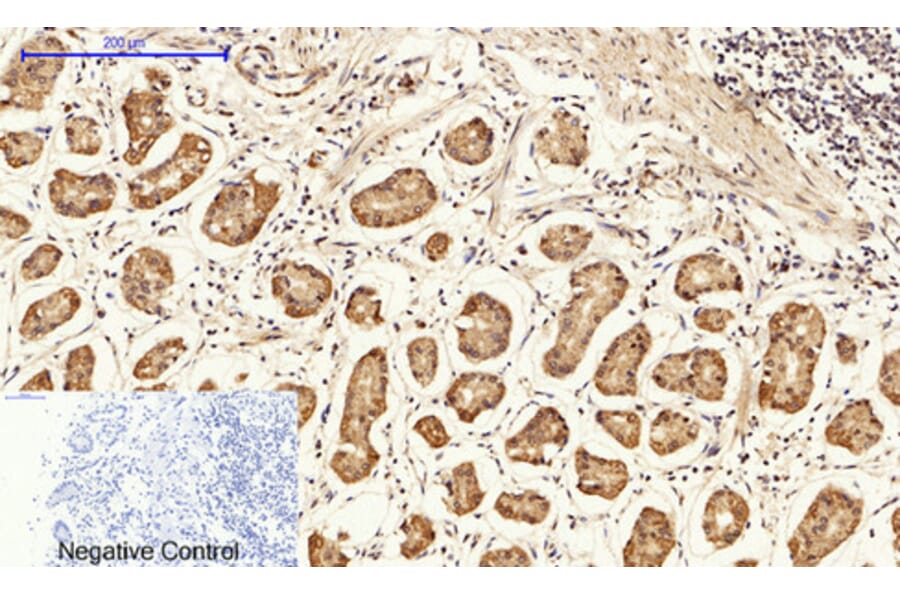
Immunohistochemical analysis of paraffin-embedded human stomach tissue using Anti-CEBP beta Antibody at 1:200 (4°C overnight). Negative control was secondary antibody only.
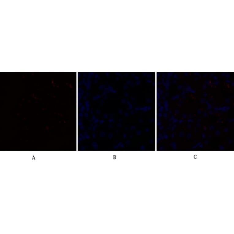
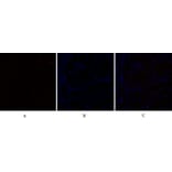
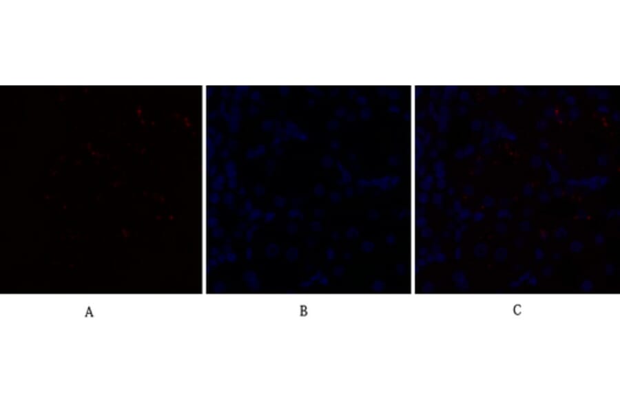
Immunofluorescence analysis of rat kidney tissue using Anti-CEBP beta Antibody (red) at 1:200 (4°C overnight). Cy3 labelled secondary antibody was used at 1:300 (RT 50min). Panel A: Target. Panel B: DAPI. Panel C: Merge.
Immunofluorescence analysis of human stomach tissue using Anti-CEBP beta Antibody (red) at 1:200 (4°C overnight). Cy3 labelled secondary antibody was used at 1:300 (RT 50min). Panel A: Target. Panel B: DAPI. Panel C: Merge.


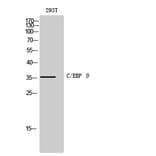

![Western Blot - Anti-CEBP Beta Antibody [ARC0017] (A308846) - Antibodies.com](https://cdn.antibodies.com/image/catalog/308/A308846_1.jpg?profile=product_alternative)