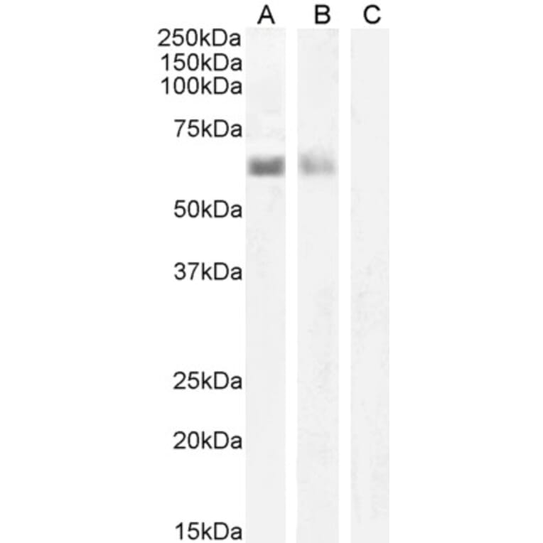
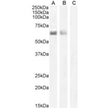
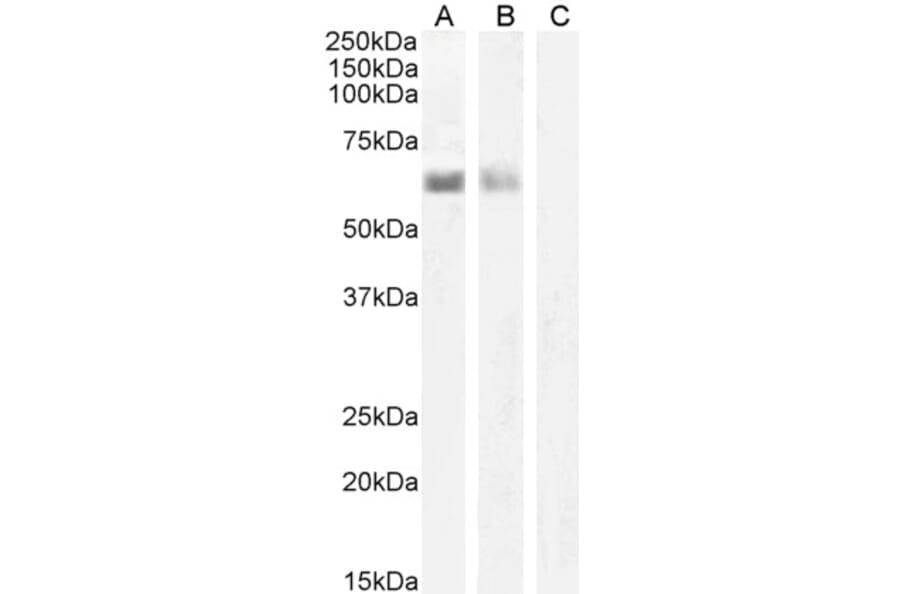
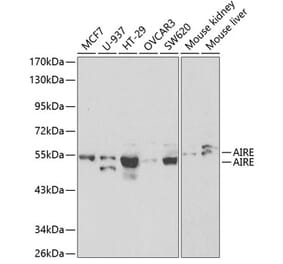
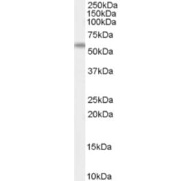
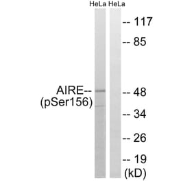
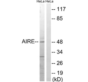
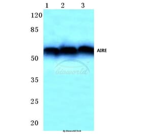
AIRE expression in nuclear Jurkat cell (A), Human Spleen (B), and negative control Human Smooth Muscle (C) lysates analyzed by western blot. Cells were lysed in RIPA buffer and 35µg protein was run per lane. Primary incubation was performed with Anti-AIRE Antibody (A82607) at 1µg/ml (A) or 2µg/ml (B-C) and detected by chemiluminescence.
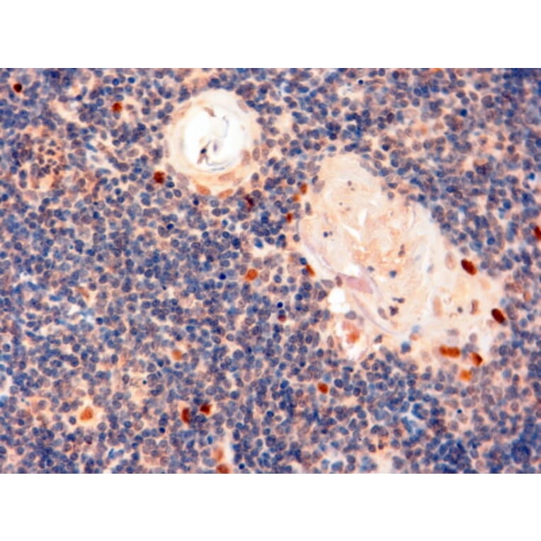
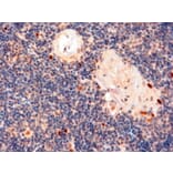
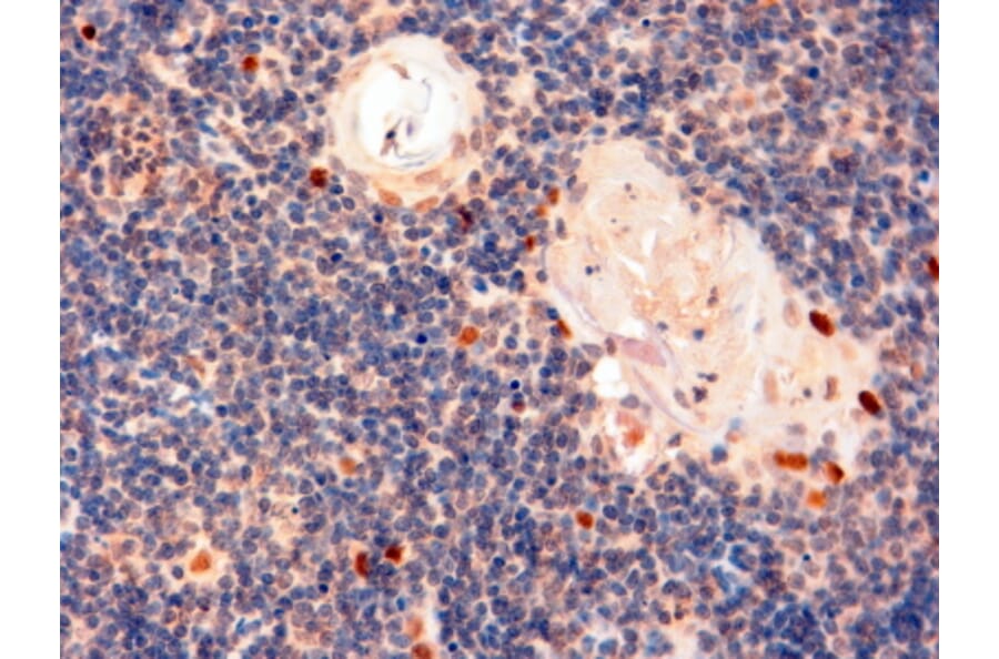
AIRE expression in Human Thymus analyzed by immunohistochemistry. Tissue was paraffin-embedded, and antigen retrieval was achieved by steaming in Tris/EDTA buffer, pH 9. Staining was performed with Anti-AIRE Antibody (A82607) at 2µg/ml and revealed with horseradish peroxidase (HRP).
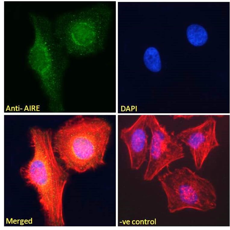
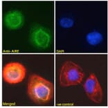
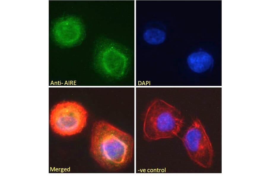
AIRE expression in U2OS cells analyzed by immunofluorescence. Cells were permeabilized with 0.15% Triton. Staining was performed with Anti-AIRE Antibody (A82607) at 10µg/ml for 1 hour and Alexa Fluor 488 secondary antibody at 2µg/ml. Nuclear membrane and nucleoplasm staining shown and nuclei were stained with DAPI (blue) while actin filaments were stained with phalloidin (red). Negative control: Goat IgG Isotype Control at 10µg/ml followed by Alexa Fluor 488 secondary antibody at 2µg/ml.
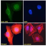
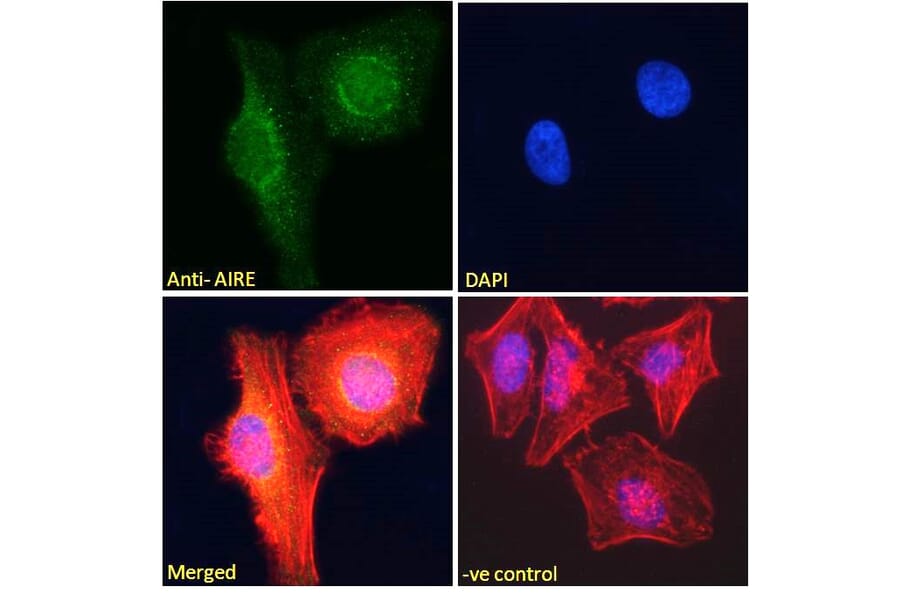
AIRE expression in HeLa cells analyzed by immunofluorescence. Cells were permeabilized with 0.15% Triton. Staining was performed with Anti-AIRE Antibody (A82607) at 10µg/ml for 1 hour and Alexa Fluor 488 secondary antibody at 2µg/ml. Nuclear membrane and nucleoplasm staining shown and nuclei were stained with DAPI (blue) while actin filaments were stained with phalloidin (red). Negative control: Goat IgG Isotype Control at 10µg/ml followed by Alexa Fluor 488 secondary antibody at 2µg/ml.
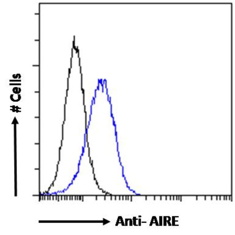
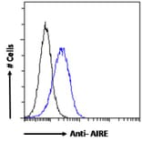
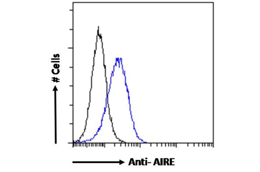
AIRE expression in Jurkat cells (blue line) analyzed by flow cytometry. Cells were fixed in PFA and permeabilized with 0.5% Triton. Staining was performed with Anti-AIRE Antibody (A82607) at 10µg/ml overnight and Alexa Fluor 488 secondary antibody at 1µg/ml. Negative Control: Goat IgG Isotype Control (black line) followed by Alexa Fluor 488 secondary antibody.