Supplied in Phosphate Buffered Saline with 50% Glycerol and 5mM Sodium Azide.
Storage
Shipped at 4°C. Upon delivery aliquot and store at -20°C. Avoid freeze/thaw cycles.
General Notes
This antibody can be used to verify the expression, size, and stability of both AcGFP and eGFP fusion proteins in western blotting experiments, and to amplify GFP signals in tissues of transgenic animals.
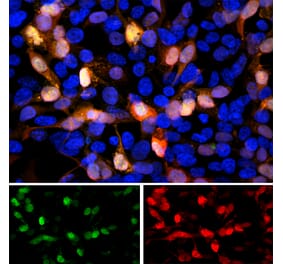
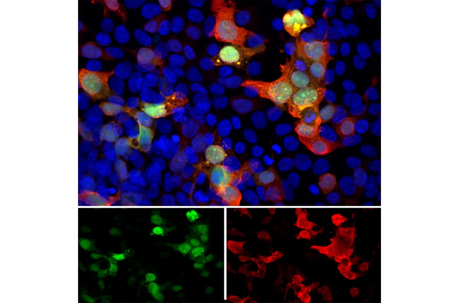
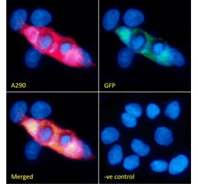
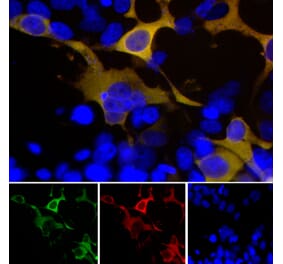
Immunofluorescent analysis of transfected HEK293 cells with a GFP-construct in green stained with Anti-GFP Antibody (A104347), at a dilution of 1:5,000, in red. The blue is Hoechst staining of nuclear DNA. Anti-GFP Antibody (A104347) reveals GFP protein expressed only in transfected cells; as a result transfected cells express both red and green signals and appear an orange-golden color. Untransfected cells show neither signal.
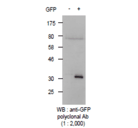
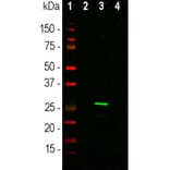
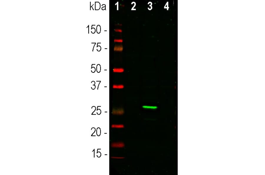
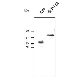
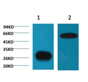
Western blot analysis of HEK293 cell lysates using Anti-GFP Antibody (A104347), at a dilution of 1:1,000, in green. The lanes contain: [Lane 1] protein standard, [Lane 2] non-transfected control cells, [Lane 3] cells transfected with a GFP construct, and [Lane 4] cells transfected with an mCherry construct. Strong band at ~27kDa corresponds to GFP protein detected only in cells transfected with GFP construct. This antibody does not recognize the mCherry protein.
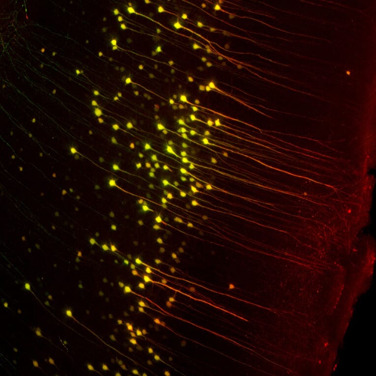
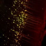
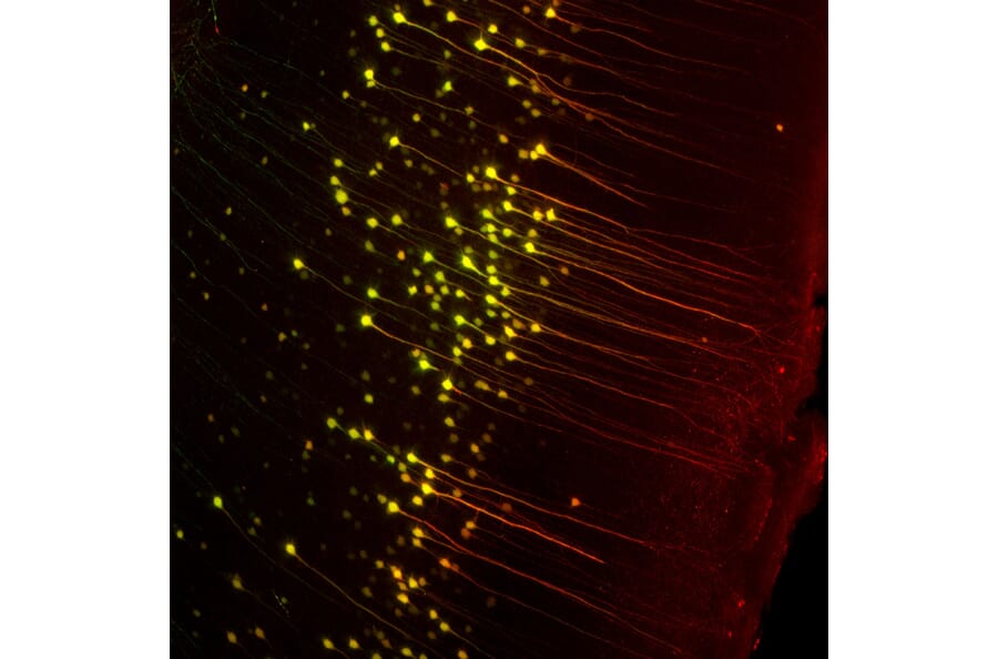
Mice transgenic for green fluorescent protein (GFP) under the Thy1 promoter express GFP in cerebral pyramidal cells. The image is of a 400µM maximum intensity optical section taken from an entire Thy1-GFP mouse brain “cleared”. Then stained the brain with Anti-GFP Antibody (A104347) coupled to a red dye. As expected, the green and red signals superimpose in the perikarya of the pyramidal cells which appear yellowish. The antibody also stains the apical dendrites which have a lower level of GFP expression, effectively amplifying the GFP signal.
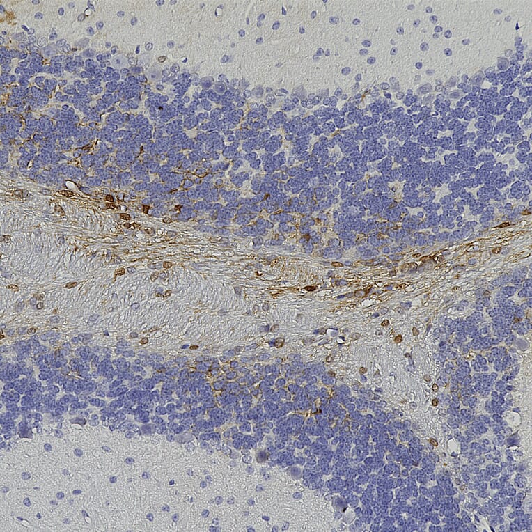
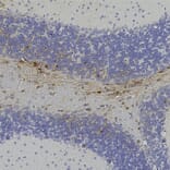
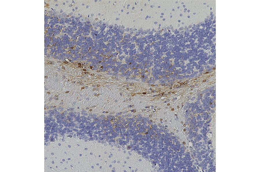
Immunohistochemistry analysis of formalin fixed paraffin embedded GFP transduced mouse brain with Anti-GFP Antibody (A104347) at a dilution of 1:5,000 detected with DAB (brown) using the Vector Elite ABC-HRP detection and reagents with citra buffer retrieval. Counterstained with Hematoxylin (blue). Anti-GFP Antibody (A104347) specifically detected GFP positive cells in the cerebellum.
Publishing research using Anti-GFP Antibody (A104347)? Please let us know so that we can list the citation on this page.


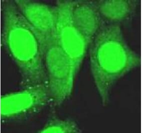
![Fluorescent image of COS1 cells due to GFP of GST-ZIPK fusion protein expressed in HEK293T cells (Right) and the same cells were immunostained using Anti-GFP Antibody [1A5], followed by Anti-Rat IgG (Texas Red) (Left). Note that fluorescence by the immunofluorescent staining using Anti-GFP Antibody [1A5] is much stronger than fluorescence due to GFP.](https://cdn.antibodies.com/image/catalog/0/A251_1.png?profile=product_alternative)