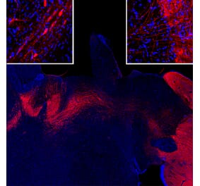
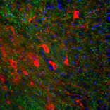
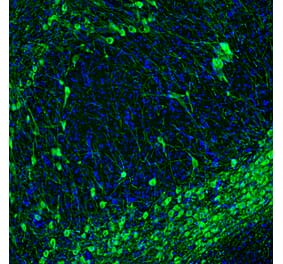
Immunofluorescent analysis of rat brain section stained with Anti-Tyrosine Hydroxylase Antibody (A104319), at a dilution of 1:10,000, in red, and co-stained with Anti-NF-H Antibody [AH1] (A85340), at a dilution of 1:1,000, in green. The blue is Hoechst staining of nuclear DNA. Following transcardial perfusion of rat with 4% paraformaldehyde, brain was post fixed for 24 hours, cut to 45µM, and free-floating sections were stained with the above antibodies. Anti-Tyrosine Hydroxylase Antibody (A104319) stains the striatal TH expressing interneurons, while Anti-NF-H Antibody [AH1] (A85340) labels axons from other neuronal cells.
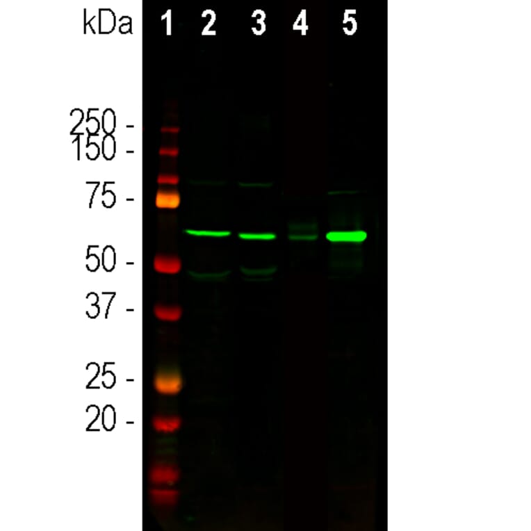
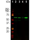
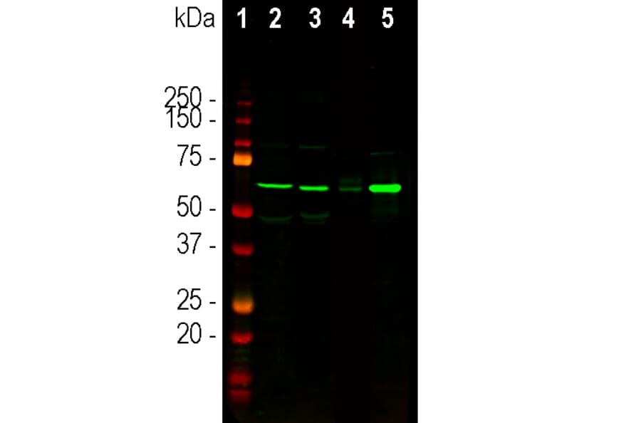
Western Blot - Anti-Tyrosine Hydroxylase Antibody (A104319)
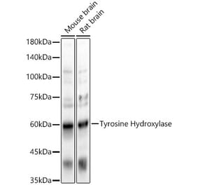
Western blot analysis of different tissue and cell lysates using Anti-Tyrosine Hydroxylase Antibody (A104319), at a dilution of 1:5,000, in green. The lanes contain: [Lane 1] protein standards (red), [Lane 2] rat brain, [Lane 3] mouse brain, [Lane 4] SH-SY5Y cells, and [Lane 5] PC12 cells. The strong band at about 60 kDa corresponds to Tyrosine Hydroxylase protein expected in brain and PC12 extracts, but not in SH-SY5Y cells.
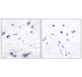
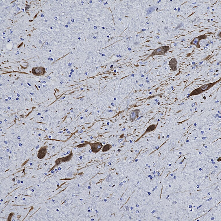
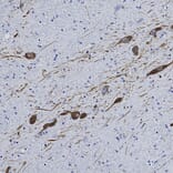
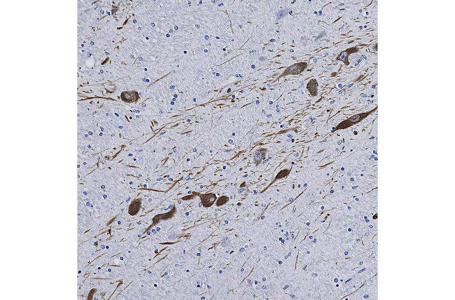
Immunohistochemistry analysis of a NBF fixed paraffin embedded human midbrain section with Anti-Tyrosine Hydroxylase Antibody (A104319) at a dilution of 1:10,000. Anti-Tyrosine Hydroxylase Antibody (A104319) labels dopaminergic neurons and their axons traversing the striatum. Note: this antibody performs well in testing with 4% PFA and standard NBF fixed mouse, rat and human tissue.
Publishing research using Anti-Tyrosine Hydroxylase Antibody (A104319)? Please let us know so that we can list the citation on this page.
Alternative products to Anti-Tyrosine Hydroxylase Antibody (A104319)


![Immunofluorescence - Anti-Tyrosine Hydroxylase Antibody [4H2] (A104315) - Antibodies.com](https://cdn.antibodies.com/image/catalog/104/A104315_1.jpg?profile=product_alternative)