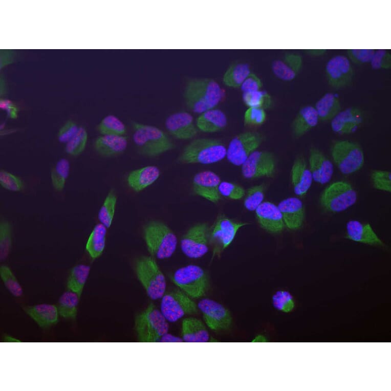
Human HeLa cells stained with Anti-SF3B4 Antibody (red), Anti-Vimentin Antibody (A85421 | green) and DNA (blue, stained with DAPI). The Anti-SF3B4 Antibody reveals strong granular nuclear staining which is a little different from the DNA stain and presumably reflects splicosomal complexes. The Anti-Vimentin Antibody stains the cytoplasmic intermediate filament network of the HeLa cells.
Immunofluorescent analysis of HeLa cells stained with Anti-SF3B4 Antibody [3A1] (A85417), at a dilution of 1:1,000, in red, and co-stained with Anti-Vimentin Antibody (A85421), at a dilution of 1:10,000, in green. The blue is DAPI staining of nuclear DNA. The Anti-SF3B4 Antibody [3A1] (A85417) reveals strong granular staining of the nuclei, while the Anti-Vimentin Antibody (A85421) specifically labels cytoplasmic intermediate filaments.
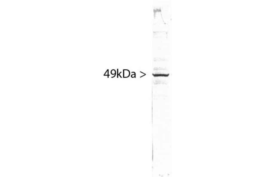
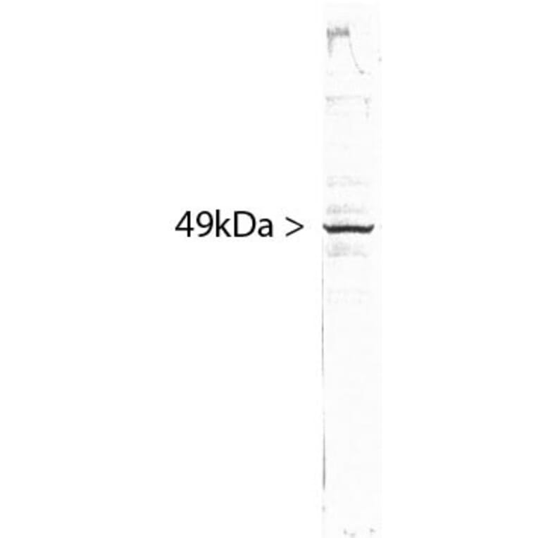
Western blot analysis of different cell lysates, cytosol or nuclear enriched fractions, using Anti-SF3B4 Antibody [3A1] (A85417), at a dilution of 1:1,000, in green. The lanes contain samples of: [1] Protein standards, in red, [2] NIH-3T3 cytosolic fraction, [3] NIH-3T3 nuclear fraction, [4] HeLa cytosolic, and [5] HeLa nuclear fractions. The strong single band at 49 kDa represents the SF3B4 protein, which is expressed exclusively in the nuclei. The same blot was simultaneously probed with Anti-GAPDH Antibody (A85377), at a dilution of 1:20,000, in red. The 37 kDa band corresponds to the GAPDH protein, detected mainly in the cytosolic fractions of these cells.


![Immunofluorescence - Anti-SF3B4 Antibody [3A1] (A85417) - Antibodies.com](https://cdn.antibodies.com/image/catalog/85/A85417_3.jpg?profile=product_top)
![Western Blot - Anti-SF3B4 Antibody [3A1] (A85417) - Antibodies.com](https://cdn.antibodies.com/image/catalog/85/A85417_4.jpg?profile=product_top)

![Immunofluorescence - Anti-SF3B4 Antibody [3A1] (A85417) - Antibodies.com](https://cdn.antibodies.com/image/catalog/85/A85417_3.jpg?profile=product_top_thumb)
![Western Blot - Anti-SF3B4 Antibody [3A1] (A85417) - Antibodies.com](https://cdn.antibodies.com/image/catalog/85/A85417_4.jpg?profile=product_top_thumb)

![Immunofluorescence - Anti-SF3B4 Antibody [3A1] (A85417) - Antibodies.com](https://cdn.antibodies.com/image/catalog/85/A85417_3.jpg?profile=product_image)
![Western Blot - Anti-SF3B4 Antibody [3A1] (A85417) - Antibodies.com](https://cdn.antibodies.com/image/catalog/85/A85417_4.jpg?profile=product_image)