Supplied in Phosphate Buffered Saline, pH 7.3, with 50% Glycerol and 0.02% Sodium Azide.
Stockage
Shipped at 4°C. Upon delivery aliquot and store at -20°C. Avoid freeze / thaw cycles.
Synonymes
40S ribosomal protein S3a, Fte 1, Fte-1, FTE1, MFTL, ribosomal protein S3A, RS3A_HUMAN, S3A, v fos transformation effector protein, v fos transformation effector protein 1, V-fos transformation effector protein
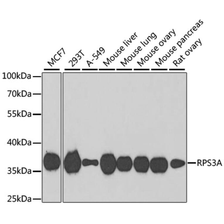
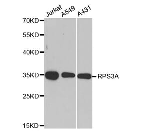
Figure 1: Western Blot - Anti-RPS3A Antibody (A15035)
Western blot analysis of extracts of various cell lines, using Anti-RPS3A Antibody (A15035) at 1:1,000 dilution. The secondary antibody was Goat Anti-Rabbit IgG H&L Antibody (HRP) at 1:10,000 dilution. Lysates/proteins were present at 25µg per lane. The blocking buffer used was 3% non-fat dry milk in TBST. Detection was with a ECL Basic Kit. Exposure time: 90s.
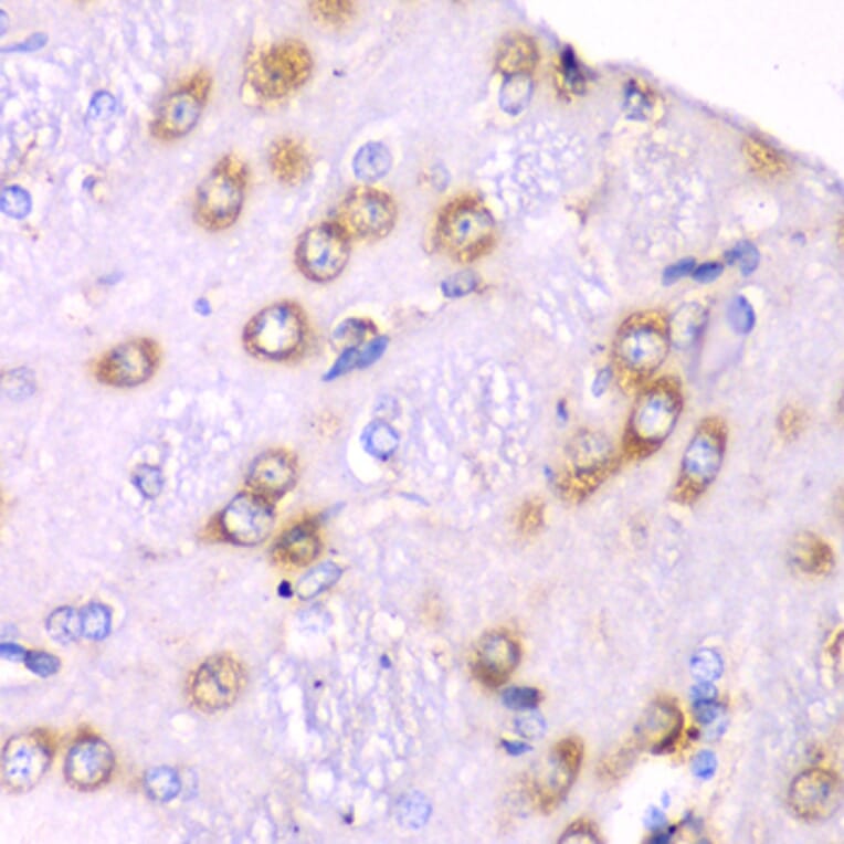
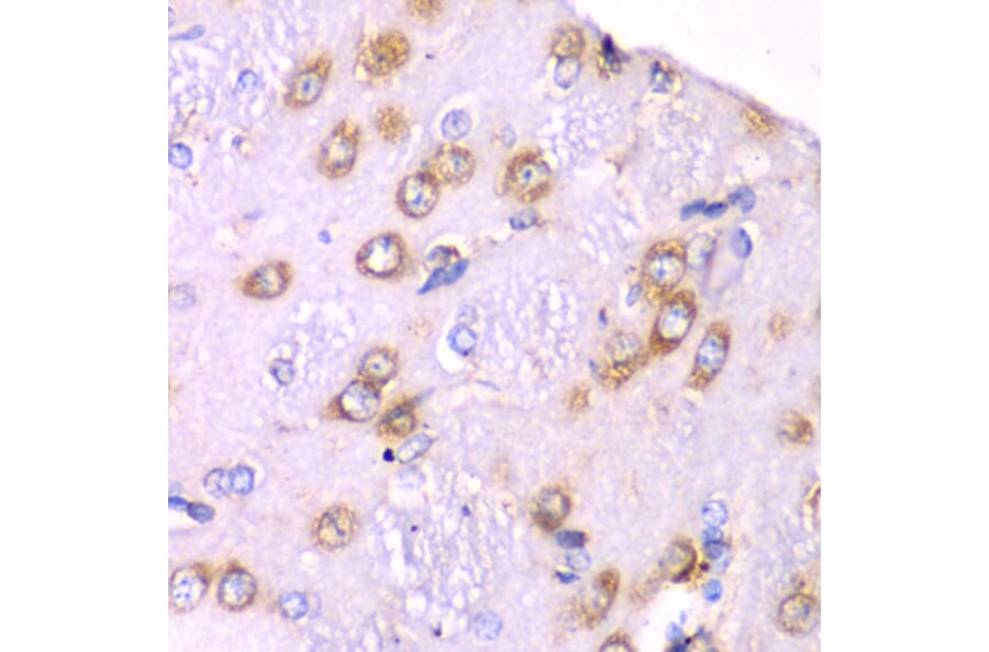
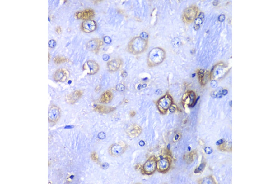
Immunohistochemistry analysis of paraffin-embedded rat brain using Anti-RPS3A Antibody (A15035) at a dilution of 1:100 (40x lens). Perform microwave antigen retrieval with 10 mM PBS buffer pH 7.2 before commencing with IHC staining protocol.
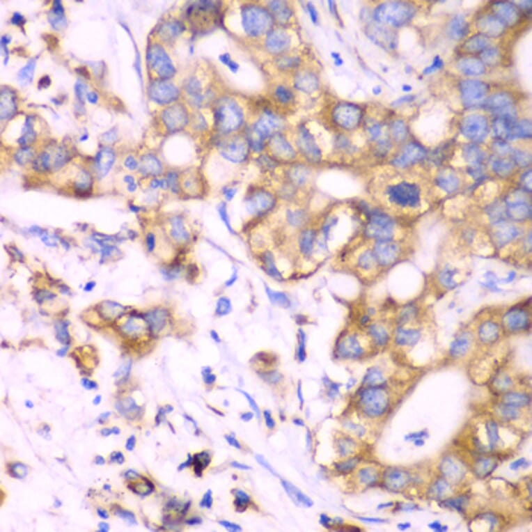
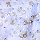
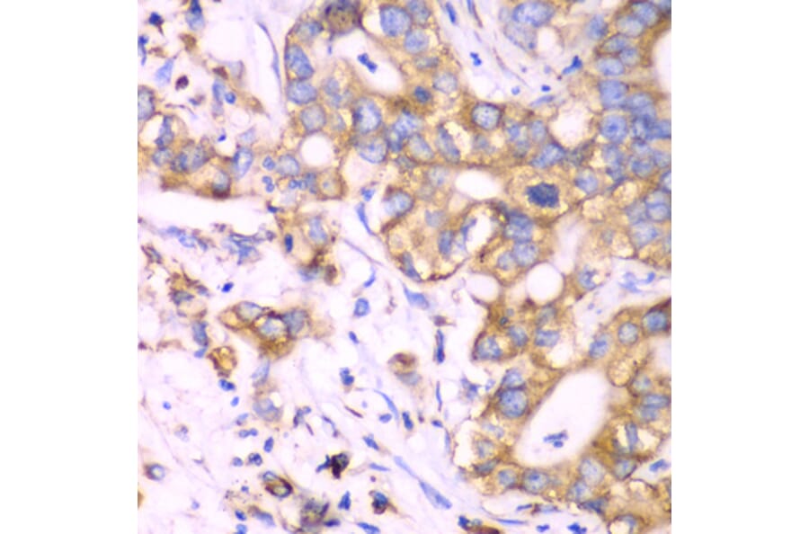
Immunohistochemistry analysis of paraffin-embedded human liver cancer using Anti-RPS3A Antibody (A15035) at a dilution of 1:100 (40x lens). Perform microwave antigen retrieval with 10 mM PBS buffer pH 7.2 before commencing with IHC staining protocol.
Immunohistochemistry analysis of paraffin-embedded mouse brain using Anti-RPS3A Antibody (A15035) at a dilution of 1:100 (40x lens). Perform microwave antigen retrieval with 10 mM PBS buffer pH 7.2 before commencing with IHC staining protocol.
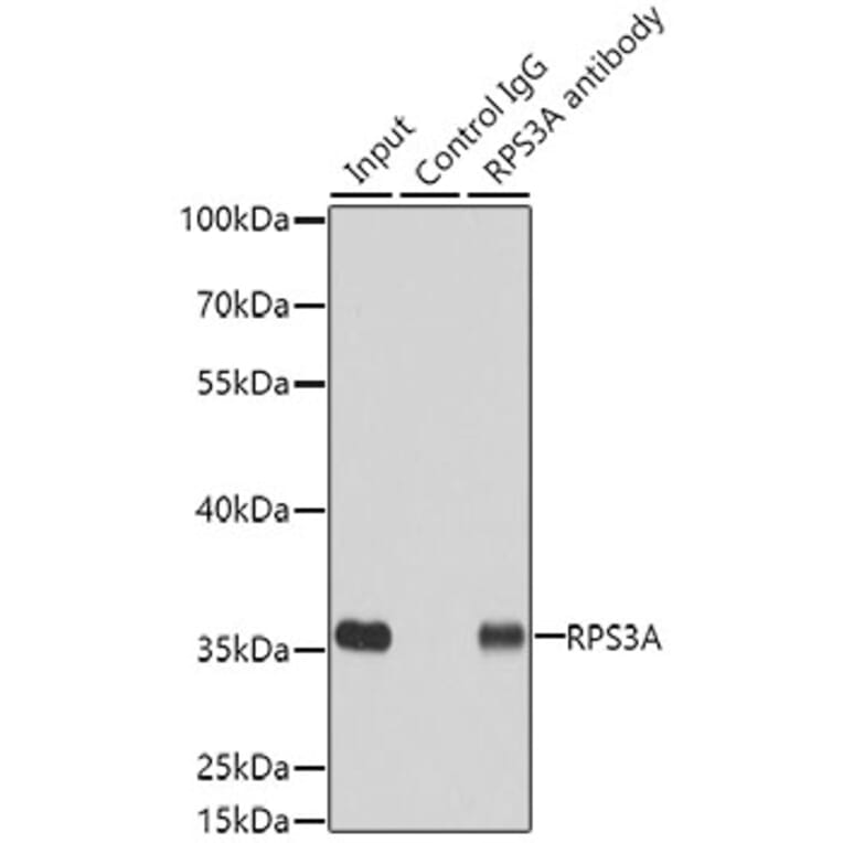
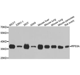
Figure 6: Western Blot - Anti-RPS3A Antibody (A15035)
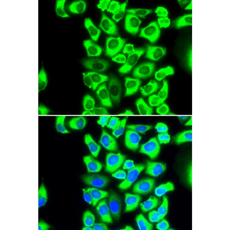
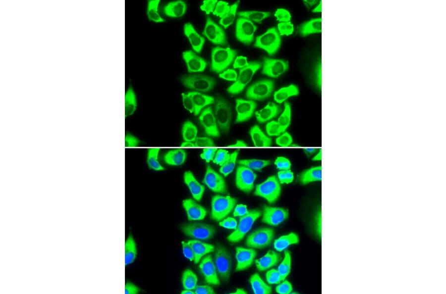
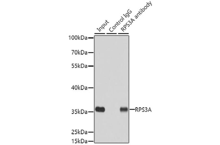
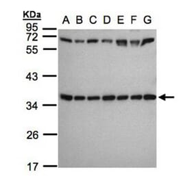
Immunoprecipitation analysis of 200µg extracts of MCF7 cells using 3µg of Anti-RPS3A Antibody (A15035). This Western blot was performed on the immunoprecipitate using Anti-RPS3A Antibody (A15035) at a dilution of 1:500.