Supplied in Phosphate Buffered Saline, pH 7.3, with 50% Glycerol and 0.05% Proclin 300.
Stockage
Shipped at 4°C. Upon delivery aliquot and store at -20°C. Avoid freeze / thaw cycles.
Synonymes
60S ribosomal protein L19, DKFZp779D216, FLJ27452, HGNC:10312, L19, MGC71997, Ribosomal Protein L19, Ribosomal protein L19, cytosolic, N terminus truncated, RL19_HUMAN
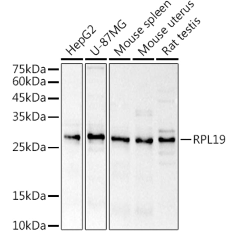
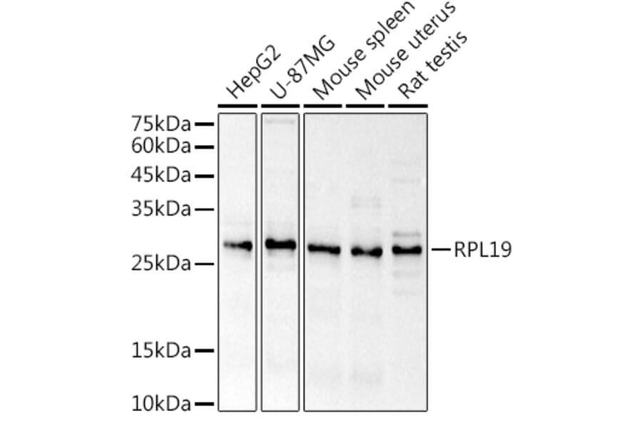
Figure 1: Western Blot - Anti-RPL19 Antibody (A307804)
Western blot analysis of extracts of various cell lines, using Anti-RPL19 Antibody (A307804) at 1:1,000 dilution. The secondary antibody was Goat Anti-Rabbit IgG H&L Antibody (HRP) at 1:10,000 dilution. Lysates/proteins were present at 25µg per lane. The blocking buffer used was 3% non-fat dry milk in TBST. Detection was with a ECL Basic Kit. Exposure time: 3s.
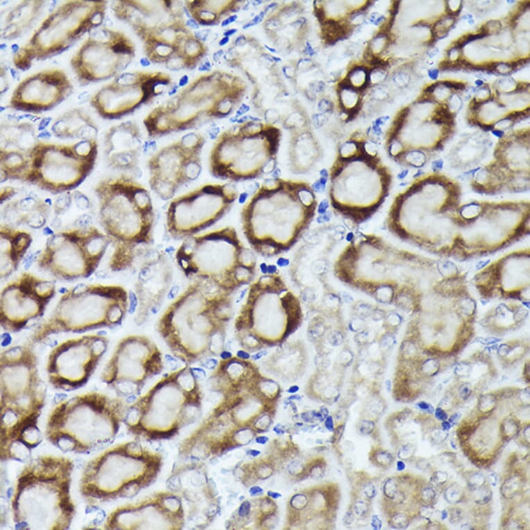
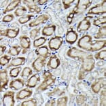
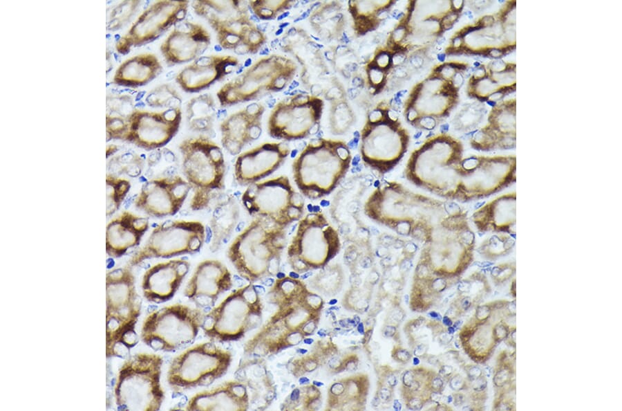
Immunohistochemistry analysis of paraffin-embedded mouse kidney using Anti-RPL19 Antibody (A307804) at a dilution of 1:200 (40x lens). Perform high pressure antigen retrieval with 10 mM citrate buffer pH 6.0 before commencing with IHC staining protocol.
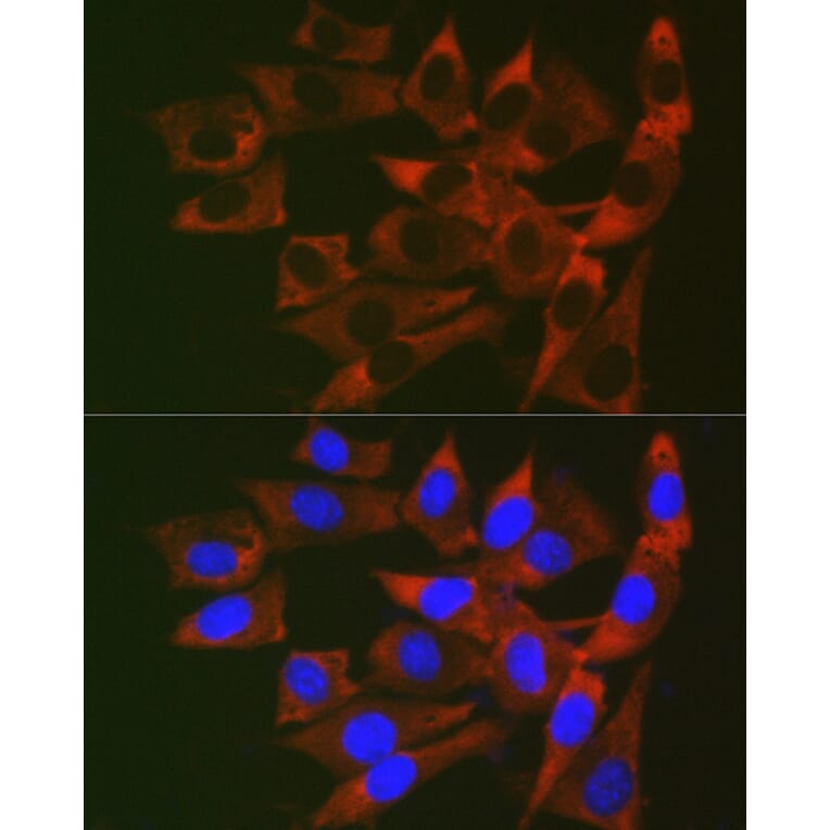
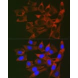
Immunofluorescence analysis of NIH/3T3 cells using Anti-RPL19 Antibody (A307804) at a dilution of 1:200 (40x lens). DAPI was used to stain the cell nuclei (blue).
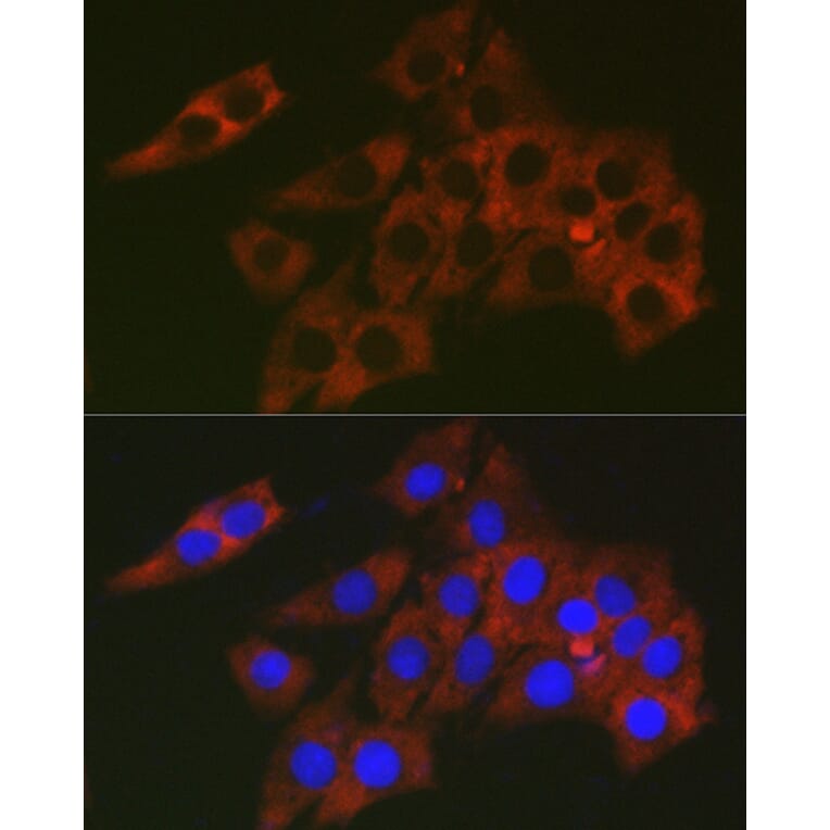
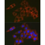
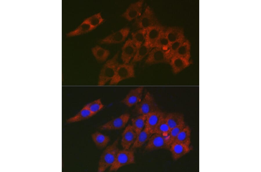
Immunofluorescence analysis of PC-12 cells using Anti-RPL19 Antibody (A307804) at a dilution of 1:200 (40x lens). DAPI was used to stain the cell nuclei (blue).
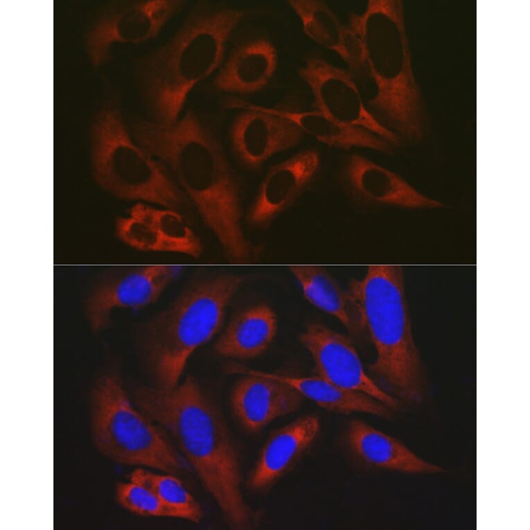
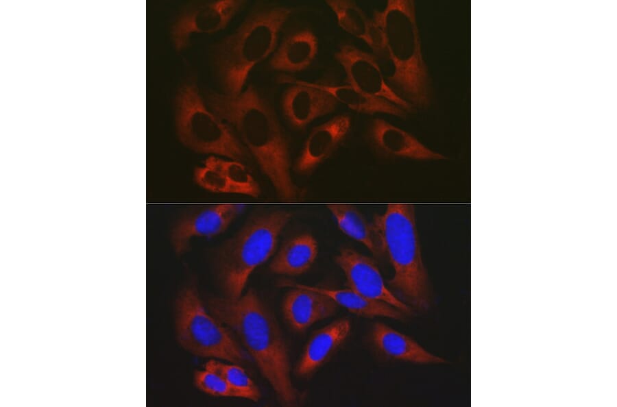
Immunofluorescence analysis of U2OS cells using Anti-RPL19 Antibody (A307804) at a dilution of 1:200 (40x lens). DAPI was used to stain the cell nuclei (blue).