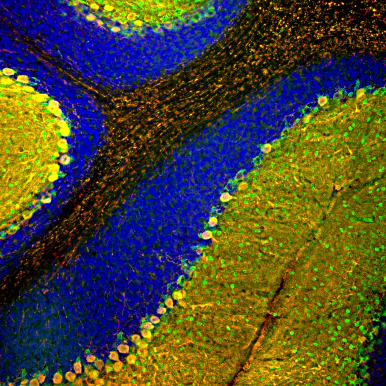
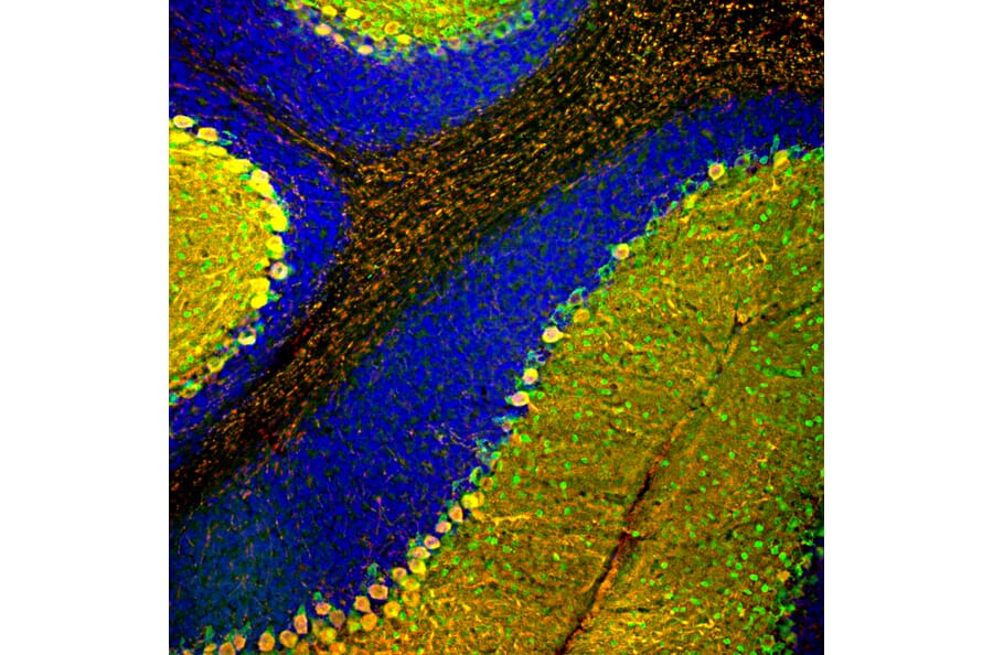
Immunofluorescent analysis of rat cerebellum section stained with Anti-Parvalbumin Antibody (1:1,000 | green) and Anti-Calbindin Antibody (A85359 | 1:2,000 | red). The blue is DAPI staining of nuclear DNA. Following transcardial perfusion of rat with 4% paraformaldehyde, brain was post fixed for 24 hours, cut to 45 µM, and free-floating sections were stained with the above antibodies. Most Purkinje cells strongly express both parvalbumin and calbindin and so appear yellow, whereas basket, stellate and Golgi cells express parvalbumin alone and so appear are green.
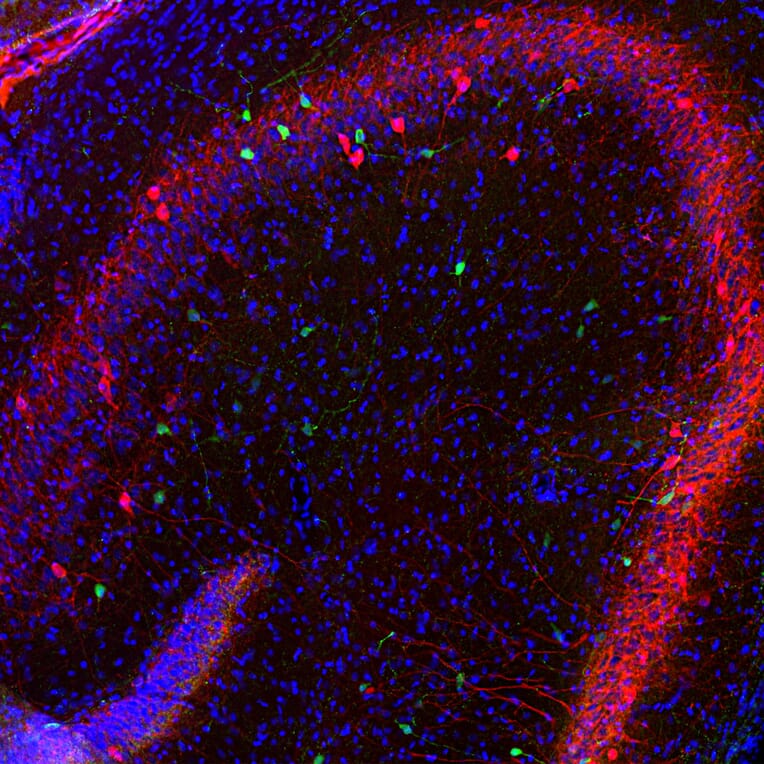
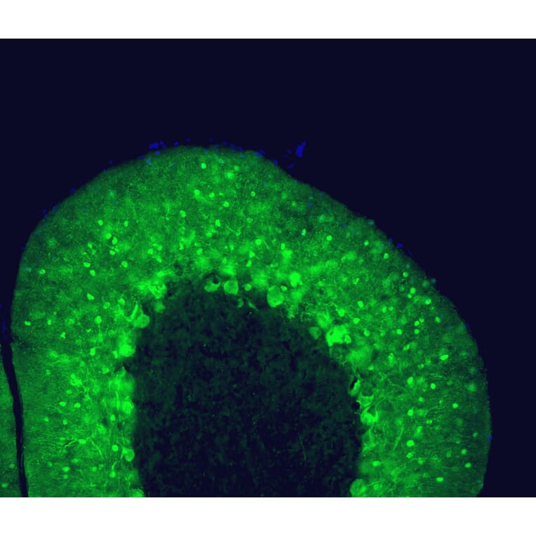
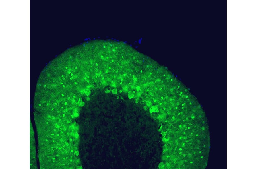
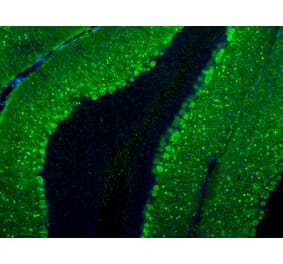
Adult rat cerebellum floating section was stained with Anti-Parvalbumin Antibody (1:1,000 | green). Parvalbumin is prominently expressed in the dendrites and perikarya of Purkinje cells and some interneurons in the molecular layer. Blue is a DNA stain.
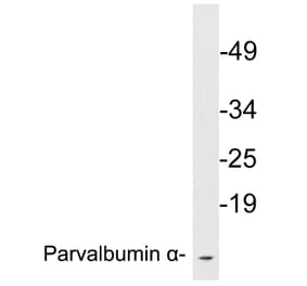
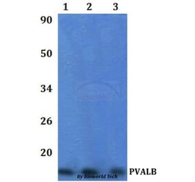
Western Blot - Anti-Parvalbumin Antibody [3C9] (A85317)
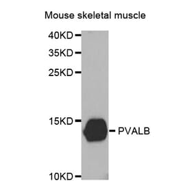
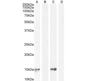
Western blot analysis of skeletal muscle lysates and His-tagged human recombinant proteins using Anti-Parvalbumin Antibody (1:1,000 | green): [1] protein standard (red), [2] mouse muscle, [3] parvalbumin, [4] calretinin, and [5] calbindin. A band at 12kDa is detected in in muscle lysate and one at 18kDa in the His-tagged recombinant parvalbumin protein lane as expected since the His-tag and other vector derived sequence adds about 6kDa to the molecule. Note that the Anti-Parvalbumin Antibody antibody is not cross-reactive with either calbindin or calretinin despite their related amino acid sequences.
Publishing research using Anti-Parvalbumin Antibody [3C9] (A85317)? Please let us know so that we can list the citation on this page.


![Immunohistochemistry - Anti-Parvalbumin Antibody [C12] (A305173) - Antibodies.com](https://cdn.antibodies.com/image/catalog/305/A305173_1.png?profile=product_alternative)
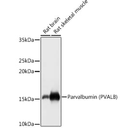
![Western Blot - Anti-Parvalbumin Antibody [ARC0385] (A307807) - Antibodies.com](https://cdn.antibodies.com/image/catalog/307/A307807_1.jpg?profile=product_alternative)