Fusion protein amino acids 597-802 (C-terminus) of rat Npas4
Host
Mouse
Clonality
Monoclonal
Clone ID
S408-79
Isotype
IgG1
Conjugate
Unconjugated
Purification
Protein G purification.
Concentration
1 mg/ml
Molecular Weight
~90 kDa
Product Form
Liquid
Formulation
Supplied in Phosphate Buffered Saline, pH 7.4, with 50% Glycerol and 0.1% Sodium Azide
Storage
Shipped at 4°C. Upon delivery aliquot and store at -20°C. Avoid freeze / thaw cycles.
Synonyms
BHLHE79, Class E basic helix-loop-helix protein 79, HLH-PAS transcription factor NXF, Neuronal PAS domain-containing protein 4, Neuronal PAS4, NXF, PAS domain-containing protein 10, PASD10
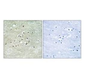
Western Blot - Anti-NPAS4 Antibody [S408-79] (A305032)
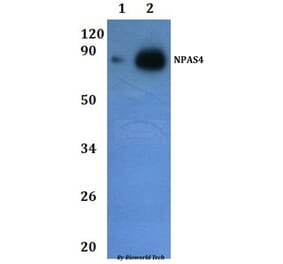
Western blot analysis of rat brain showing detection of ~90 kDa NPAS4 protein using Anti-NPAS4 Antibody [S408-79] (A305032) at 1:1,000 for 16 hours at 4°C. Lane 1: MW Ladder. Lane 2: rat Brain. Load: 20 µg. Block: 2% GE Healthcare Blocker for 1 hour at room temperature. The secondary antibody used was Goat Anti-Mouse IgG: HRP at 1:200 for 1 hour at room temperature. Color Development: ECL solution for 6 min at room temperature. Predicted/Observed Size: ~90 kDa. Other Band(s): ~60, 45, 40, 38, 25, 20 kDa.
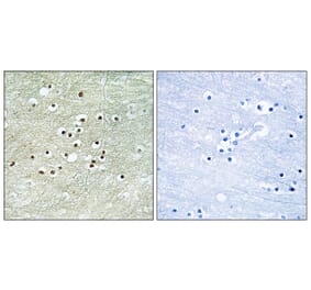
Immunocytochemistry/Immunofluorescence analysis of human neuroblastoma cell line (SK-N-BE, fixed in 4% formaldehyde for 15 min at room temperature, using Anti-NPAS4 Antibody [S408-79] (A305032), at 1:100 for 60 minutes at room temperature. The secondary antibody used was Goat Anti-Mouse ATTO 488 at 1:100 for 60 minutes at room temperature. Counterstain: Phalloidin Texas Red F-Actin stain; DAPI (blue) nuclear stain at 1:1000, 1:5,000 for 60min room temperature, 5min room temperature. Localization: Cytoplasm. Magnification: 60X.(A) DAPI (blue) nuclear stain (B) Phalloidin Texas Red F-Actin stain (C) NPAS4 Antibody (D) Composite.
Publishing research using Anti-NPAS4 Antibody [S408-79] (A305032)? Please let us know so that we can list the citation on this page.
Alternative products to Anti-NPAS4 Antibody [S408-79] (A305032)


![Western Blot - Anti-NPAS4 Antibody [S408-79] (A305032) - Antibodies.com](https://cdn.antibodies.com/image/catalog/305/A305032_1.png?profile=product_top)
![Immunocytochemistry/Immunofluorescence - Anti-NPAS4 Antibody [S408-79] (A305032) - Antibodies.com](https://cdn.antibodies.com/image/catalog/305/A305032_2.png?profile=product_top)
![Western Blot - Anti-NPAS4 Antibody [S408-79] (A305032) - Antibodies.com](https://cdn.antibodies.com/image/catalog/305/A305032_1.png?profile=product_top_thumb)
![Immunocytochemistry/Immunofluorescence - Anti-NPAS4 Antibody [S408-79] (A305032) - Antibodies.com](https://cdn.antibodies.com/image/catalog/305/A305032_2.png?profile=product_top_thumb)
![Western Blot - Anti-NPAS4 Antibody [S408-79] (A305032) - Antibodies.com](https://cdn.antibodies.com/image/catalog/305/A305032_1.png?profile=product_image)
![Immunocytochemistry/Immunofluorescence - Anti-NPAS4 Antibody [S408-79] (A305032) - Antibodies.com](https://cdn.antibodies.com/image/catalog/305/A305032_2.png?profile=product_image)