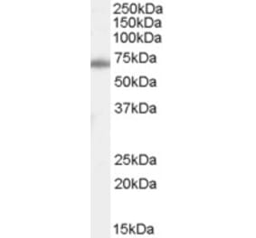
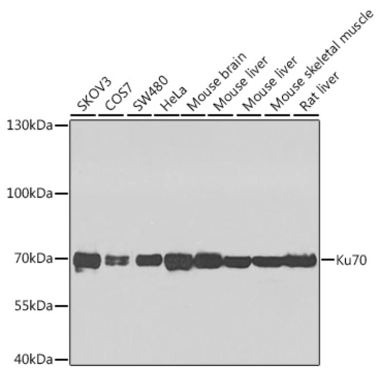
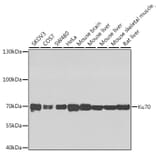
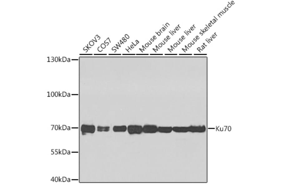
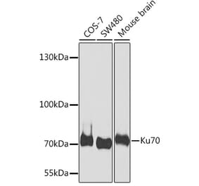
Western blot analysis of extracts of various cell lines, using Anti-Ku70 Antibody (A16783) at 1:1,000 dilution. The secondary antibody was Goat Anti-Rabbit IgG H&L Antibody (HRP) at 1:10,000 dilution. Lysates/proteins were present at 25µg per lane. The blocking buffer used was 3% non-fat dry milk in TBST. Detection was with a ECL Enhanced Kit (RM00021). Exposure time: 60s.
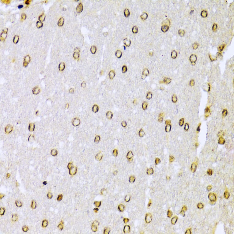
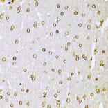
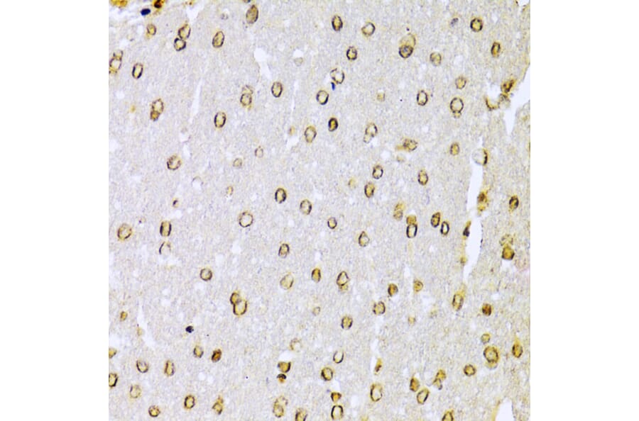
Immunohistochemistry analysis of paraffin-embedded rat brain using Anti-Ku70 Antibody (A16783) at a dilution of 1:100 (40x lens). Perform microwave antigen retrieval with 10 mM PBS buffer pH 7.2 before commencing with IHC staining protocol.
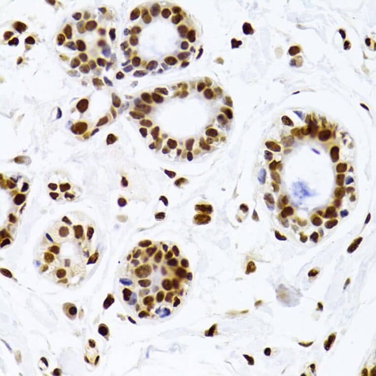
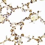
Immunohistochemistry analysis of paraffin-embedded human breast tissue using Anti-Ku70 Antibody (A16783) at a dilution of 1:100 (40x lens). Perform microwave antigen retrieval with 10 mM PBS buffer pH 7.2 before commencing with IHC staining protocol.
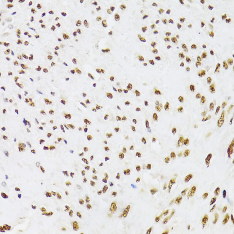
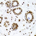
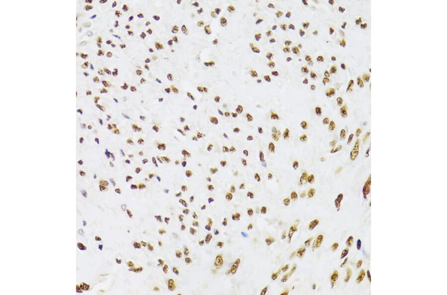
Immunohistochemistry analysis of paraffin-embedded human leiomyoma of uterus using Anti-Ku70 Antibody (A16783) at a dilution of 1:100 (40x lens). Perform microwave antigen retrieval with 10 mM PBS buffer pH 7.2 before commencing with IHC staining protocol.
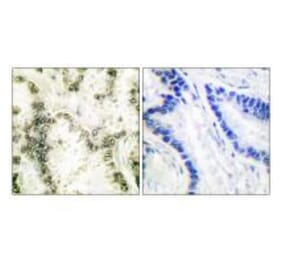
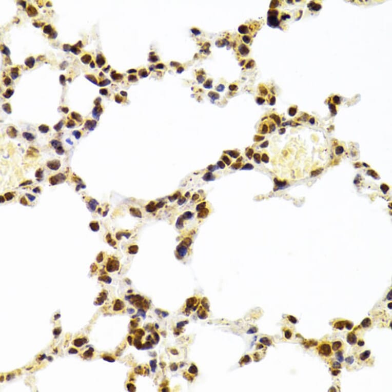
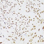
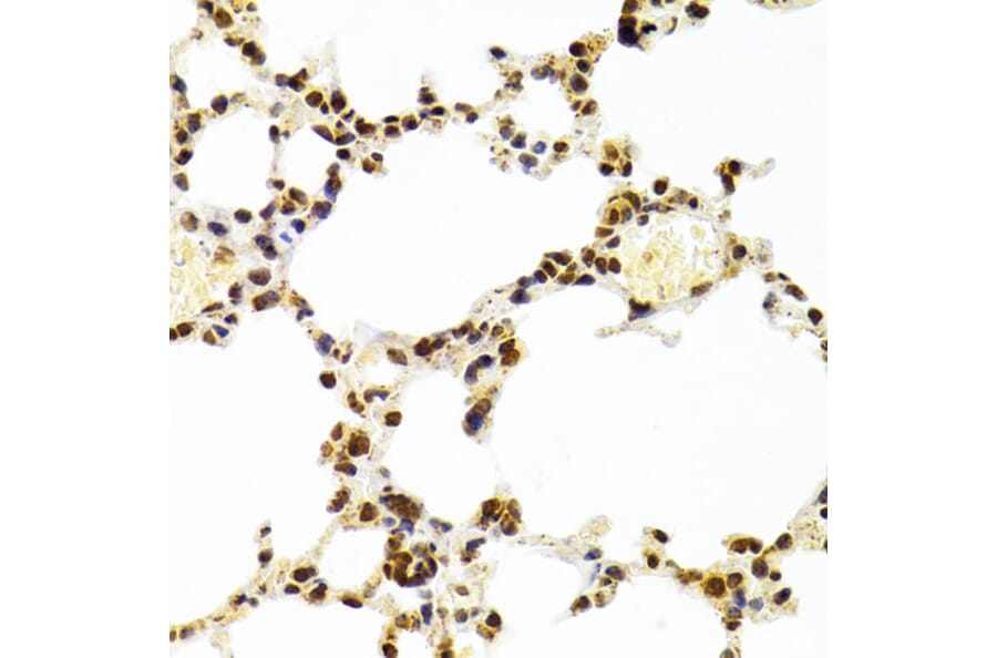
Immunohistochemistry analysis of paraffin-embedded mouse lung using Anti-Ku70 Antibody (A16783) at a dilution of 1:100 (40x lens). Perform microwave antigen retrieval with 10 mM PBS buffer pH 7.2 before commencing with IHC staining protocol.
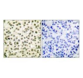
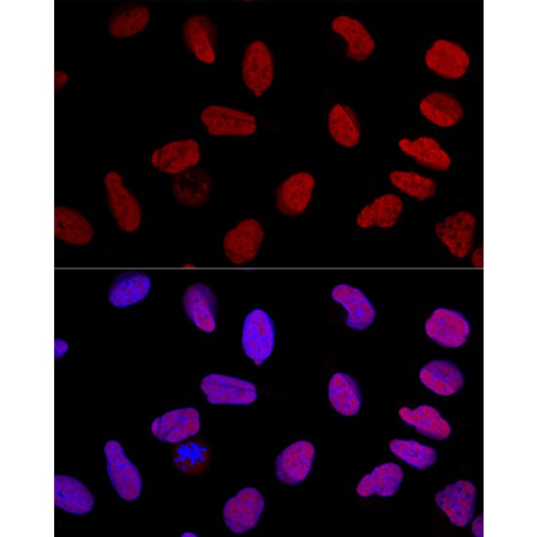
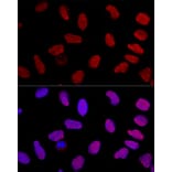
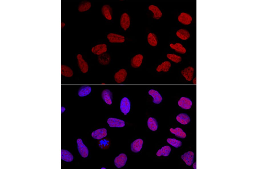
Confocal immunofluorescence analysis of U-2 OS cells using Anti-Ku70 Antibody (A16783) at a dilution of 1:100. DAPI was used to stain the cell nuclei (blue).


![Western Blot - Anti-Ku70 Antibody [ARC0551] (A80789) - Antibodies.com](https://cdn.antibodies.com/image/catalog/80/A80789_1.jpg?profile=product_alternative)
![Flow Cytometry - Anti-Ku70 Antibody [KU729] (A248673) - Antibodies.com](https://cdn.antibodies.com/image/catalog/248/A248673_1.jpg?profile=product_alternative)
![Flow Cytometry - Anti-Ku70 Antibody [KU729] - BSA and Azide free (A251855) - Antibodies.com](https://cdn.antibodies.com/image/catalog/251/A251855_1.jpg?profile=product_alternative)