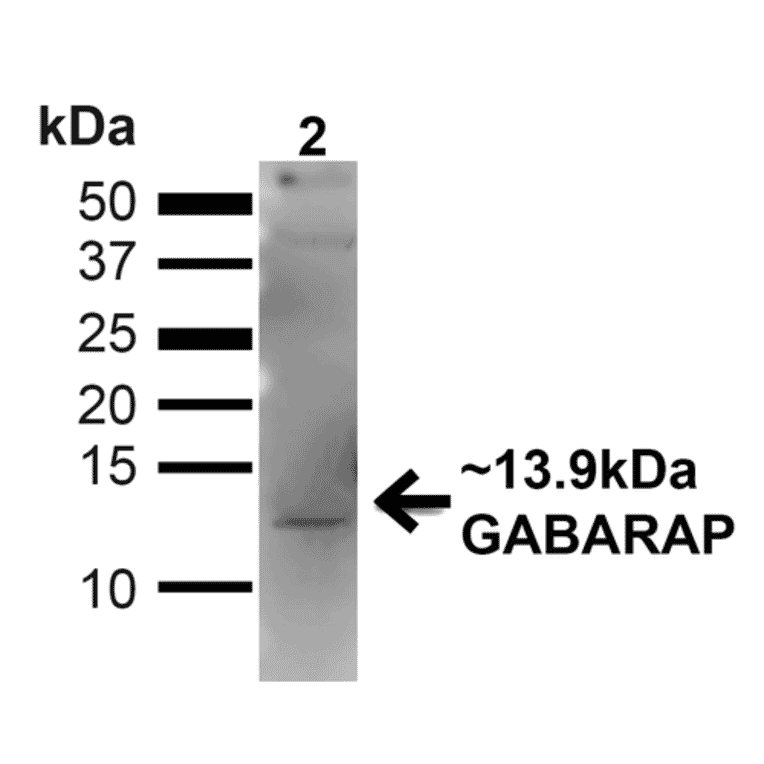
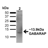
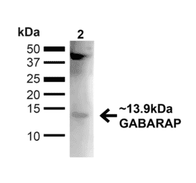
Western blot analysis of mouse kidney showing detection of ~13.9kDa GABARAP protein using Anti-GABARAP Antibody (A304934) at 1:1,000 for 1 hour at room temperature. Lane 1: MW Ladder. Lane 2: mouse kidney (20 µg). Load: 20 µg. Block: 5% milk + TBST for 1 hour at room temperature. The secondary antibody used was Goat Anti-Rabbit: HRP at 1:2000 for 1 hour at room temperature. Color Development: TMB solution for 12 min at room temperature. Predicted/Observed Size: ~13.9kDa. Other Band(s): ~40kDa (Complex).
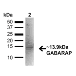
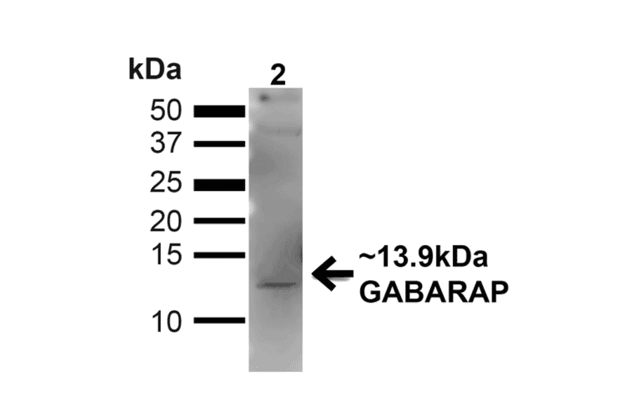
Western blot analysis of human Embryonic kidney epithelial cell line (HEK293T) lysate showing detection of ~13.9kDa GABARAP protein using Anti-GABARAP Antibody (A304934) at 1:1,000 for 1 hour at room temperature. Lane 1: MW Ladder. Lane 2: human 293T (20 µg). Load: 20 µg. Block: 5% milk + TBST for 1 hour at room temperature. The secondary antibody used was Goat Anti-Rabbit: HRP at 1:2000 for 1 hour at room temperature. Color Development: TMB solution for 12 min at room temperature. Predicted/Observed Size: ~13.9kDa. Other Band(s): ~40kDa (Complex).
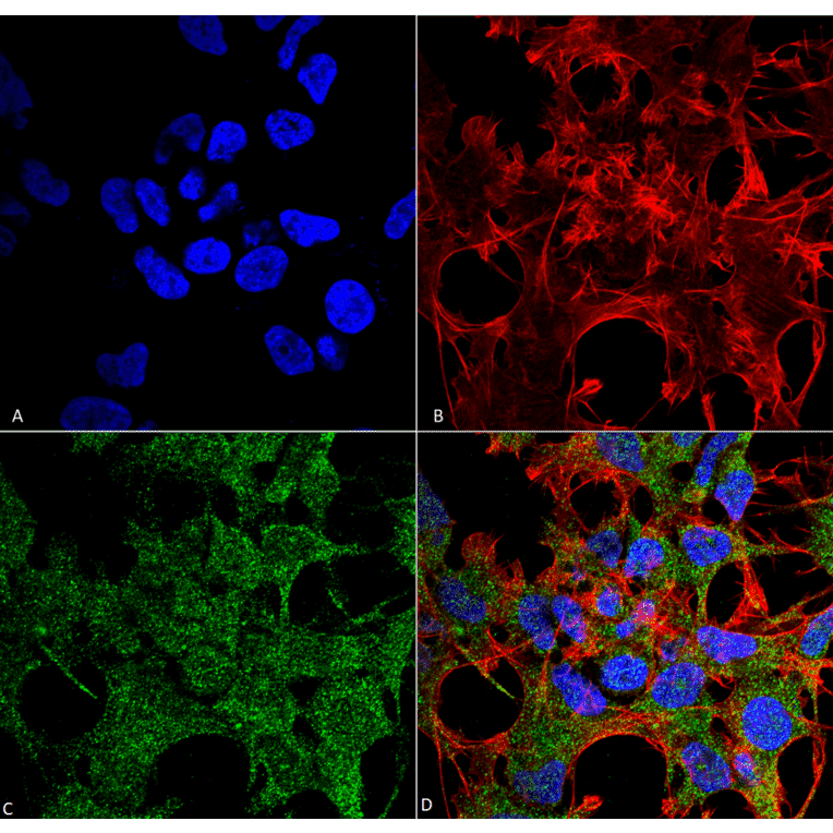
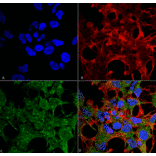
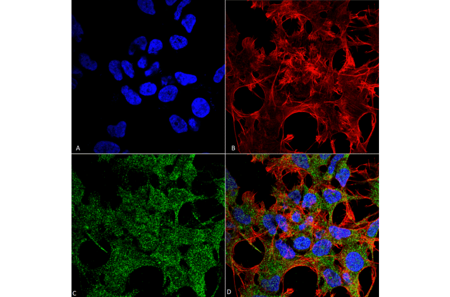
Immunocytochemistry/Immunofluorescence analysis of human neuroblastoma cell line (SK-N-BE, fixed in 4% formaldehyde for 15 min at room temperature, using Anti-GABARAP Antibody (A304934), at 1:100 for 60 minutes at room temperature. The secondary antibody used was Goat Anti-Rabbit ATTO 488 at 1:100 for 60 minutes at room temperature. Counterstain: Phalloidin Texas Red F-Actin stain; DAPI (blue) nuclear stain at 1:1000, 1:5,000 for 60min room temperature, 5min room temperature. Localization: Endomembrane System, Cytoplasm, Golgi Apparatus Membrane, Cytoplasmic Vesicle. Magnification: 60X.(A) DAPI (blue) nuclear stain (B) Phalloidin Texas Red F-Actin stain (C) GABARAP Antibody (D) Composite.
Publishing research using Anti-GABARAP Antibody (A304934)? Please let us know so that we can list the citation on this page.
Alternative products to Anti-GABARAP Antibody (A304934)


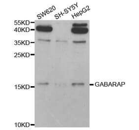
![Western Blot - Anti-GABARAP Antibody [ARC1057] (A307456) - Antibodies.com](https://cdn.antibodies.com/image/catalog/307/A307456_1.jpg?profile=product_alternative)