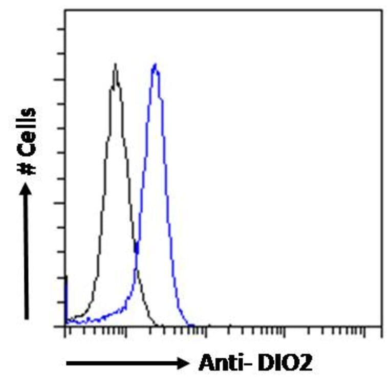
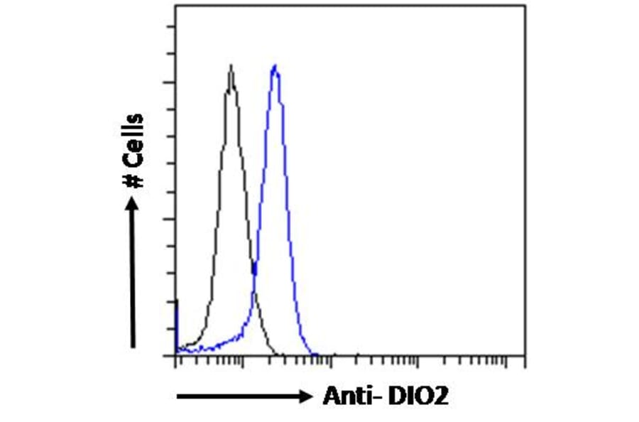
DIO2 expression in MCF7 cells (blue line) analyzed by flow cytometry. Cells were fixed in PFA and permeabilized with 0.5% Triton. Staining was performed with Anti-DIO2 Antibody (A83378) at 10µg/ml for 1 hour and Alexa Fluor 488 secondary antibody at 1µg/ml. Negative Control: Goat IgG Isotype Control (black line) followed by Alexa Fluor 488 secondary antibody.
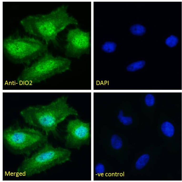
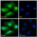
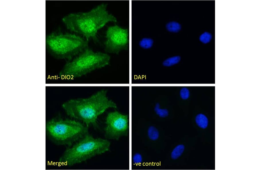
DIO2 expression in HeLa cells analyzed by immunofluorescence. Cells were permeabilized with 0.15% Triton. Staining was performed with Anti-DIO2 Antibody (A83378) at 10µg/ml for 1 hour and Alexa Fluor 488 secondary antibody at 2µg/ml. Cytoplasmic and nuclear staining shown and nuclei were stained with DAPI (blue). Negative control: Goat IgG Isotype Control at 10µg/ml followed by Alexa Fluor 488 secondary antibody at 2µg/ml.
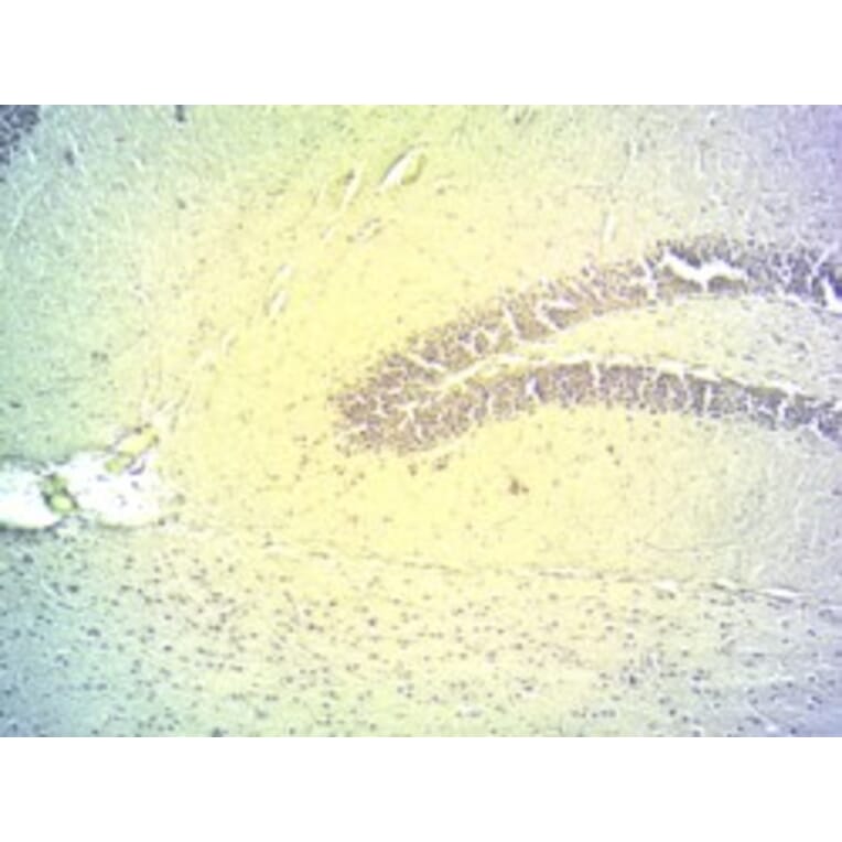
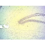
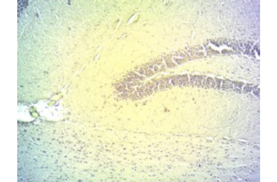
DIO2 expression in Mouse Hippocampus analyzed by immunohistochemistry. Tissue was cryosectioned after PFA perfusion fixation, and antigen retrieval was achieved by microwaving in citrate buffer, pH 4.5. Staining was performed with Anti-DIO2 Antibody (A83378) at 2µg/ml and revealed with horseradish peroxidase (HRP).
Publishing research using Anti-DIO2 Antibody (A83378)? Please let us know so that we can list the citation on this page.
Alternative products to Anti-DIO2 Antibody (A83378)