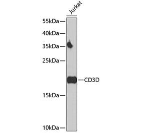
Western Blot - Anti-CD3D Antibody [ARC1741] (A308414)
Western blot analysis of extracts of Jurkat cells, using Anti-CD3D Antibody [ARC1741] (A308414) at 1:500 dilution. The secondary antibody was Goat Anti-Rabbit IgG H&L Antibody (HRP) at 1:10,000 dilution. Lysates/proteins were present at 25µg per lane. The blocking buffer used was 3% non-fat dry milk in TBST. Detection was with a ECL Basic Kit. Exposure time: 60s.
Immunofluorescence analysis of rat spleen using Anti-CD3D Antibody [ARC1741] (A308414) at a dilution of 1:100 (40x lens). DAPI was used to stain the cell nuclei (blue).
Immunofluorescence analysis of mouse spleen using Anti-CD3D Antibody [ARC1741] (A308414) at a dilution of 1:100 (40x lens). DAPI was used to stain the cell nuclei (blue).
Flow cytometry analysis of Jurkat cells, stained with Rabbit IgG isotype control (2.5 µg/ml, blue line) or Anti-CD3D Antibody [ARC1741] (A308414), (2.5 µg/ml orange line), followed by goat anti-Rabbit polyclonal antibody FITC (1:200 dilution) staining. Non-fluorescently stained Jurkat cells was used as blank control (red line).
Flow cytometry analysis of Raji cells, stained with Rabbit IgG isotype control (2.5 µg/ml, blue line) or Anti-CD3D Antibody [ARC1741] (A308414), (2.5 µg/ml orange line), followed by goat anti-Rabbit polyclonal antibody FITC (1:200 dilution) staining. Non-fluorescently stained Raji cells was used as blank control (red line).


![Western Blot - Anti-CD3D Antibody [ARC1741] (A308414) - Antibodies.com](https://cdn.antibodies.com/image/catalog/308/A308414_1.jpg?profile=product_top)
![Immunofluorescence - Anti-CD3D Antibody [ARC1741] (A308414) - Antibodies.com](https://cdn.antibodies.com/image/catalog/308/A308414_2.jpg?profile=product_top)
![Immunofluorescence - Anti-CD3D Antibody [ARC1741] (A308414) - Antibodies.com](https://cdn.antibodies.com/image/catalog/308/A308414_3.jpg?profile=product_top)
![Flow Cytometry - Anti-CD3D Antibody [ARC1741] (A308414) - Antibodies.com](https://cdn.antibodies.com/image/catalog/308/A308414_4.jpg?profile=product_top)
![Flow Cytometry - Anti-CD3D Antibody [ARC1741] (A308414) - Antibodies.com](https://cdn.antibodies.com/image/catalog/308/A308414_5.jpg?profile=product_top)
![Western Blot - Anti-CD3D Antibody [ARC1741] (A308414) - Antibodies.com](https://cdn.antibodies.com/image/catalog/308/A308414_1.jpg?profile=product_top_thumb)
![Immunofluorescence - Anti-CD3D Antibody [ARC1741] (A308414) - Antibodies.com](https://cdn.antibodies.com/image/catalog/308/A308414_2.jpg?profile=product_top_thumb)
![Immunofluorescence - Anti-CD3D Antibody [ARC1741] (A308414) - Antibodies.com](https://cdn.antibodies.com/image/catalog/308/A308414_3.jpg?profile=product_top_thumb)
![Flow Cytometry - Anti-CD3D Antibody [ARC1741] (A308414) - Antibodies.com](https://cdn.antibodies.com/image/catalog/308/A308414_4.jpg?profile=product_top_thumb)
![Flow Cytometry - Anti-CD3D Antibody [ARC1741] (A308414) - Antibodies.com](https://cdn.antibodies.com/image/catalog/308/A308414_5.jpg?profile=product_top_thumb)
![Western Blot - Anti-CD3D Antibody [ARC1741] (A308414) - Antibodies.com](https://cdn.antibodies.com/image/catalog/308/A308414_1.jpg?profile=product_image)
![Immunofluorescence - Anti-CD3D Antibody [ARC1741] (A308414) - Antibodies.com](https://cdn.antibodies.com/image/catalog/308/A308414_2.jpg?profile=product_image)
![Immunofluorescence - Anti-CD3D Antibody [ARC1741] (A308414) - Antibodies.com](https://cdn.antibodies.com/image/catalog/308/A308414_3.jpg?profile=product_image)
![Flow Cytometry - Anti-CD3D Antibody [ARC1741] (A308414) - Antibodies.com](https://cdn.antibodies.com/image/catalog/308/A308414_4.jpg?profile=product_image)
![Flow Cytometry - Anti-CD3D Antibody [ARC1741] (A308414) - Antibodies.com](https://cdn.antibodies.com/image/catalog/308/A308414_5.jpg?profile=product_image)
![Mass Cytometry - Anti-CD3 Antibody [UCHT1] (A86521) - Antibodies.com](https://cdn.antibodies.com/image/catalog/86/A86522_697.jpg?profile=product_alternative)

![Mass Cytometry - Anti-CD3 Antibody [MEM-57] (A86065) - Antibodies.com](https://cdn.antibodies.com/image/catalog/86/A86066_410.jpg?profile=product_alternative)
![Mass Cytometry - Anti-CD3 Antibody [UCHT1] - Low endotoxin, Azide free (A86528) - Antibodies.com](https://cdn.antibodies.com/image/catalog/86/A86529_702.jpg?profile=product_alternative)
![Immunohistochemistry - Anti-CD3 Antibody [RM344] (A121367) - Antibodies.com](https://cdn.antibodies.com/image/catalog/121/A121411_1.png?profile=product_alternative)
![Flow Cytometry - Anti-CD3 Antibody [TB3] (A121786) - Antibodies.com](https://cdn.antibodies.com/image/catalog/121/A121789_1.jpg?profile=product_alternative)