Synthetic peptide corresponding to the internal region (near N terminus) of human Calnexin.
Séquence
C-SKTPELNLDQFHDKT
Hôte
Goat
Clonalité
Polyclonal
Isotype
IgG
Conjuguer
Unconjugated
Purification
Purified from goat serum by ammonium sulphate precipitation followed by antigen affinity chromatography using the immunizing peptide.
Concentration
500 µg/ml
Masse moléculaire
90-100 kDa
MW prédit
67.6kDa
Forme du produit
Liquid
Formulation
Supplied in Tris Buffered Saline, pH 7.3, with 0.5% BSA and 0.02% Sodium Azide.
Stockage
Shipped at 4°C. Upon delivery aliquot and store at -20°C. Avoid freeze/thaw cycles.
Synonymes
CALX_HUMAN, CANX, CNX, FLJ26570, Histocompatibility complex class I antigen binding protein p88, IP90, Major histocompatibility complex class I antigen-binding protein p88, p90
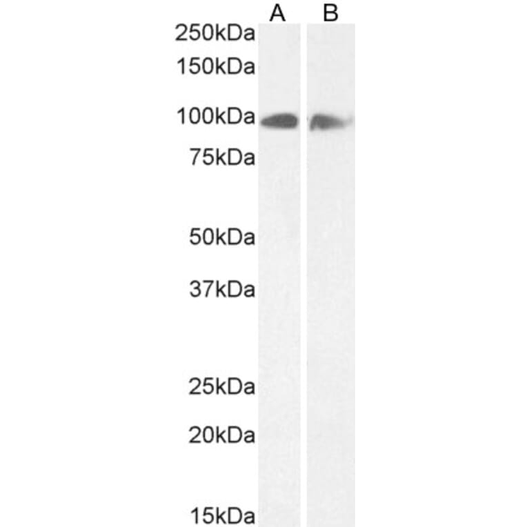
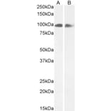
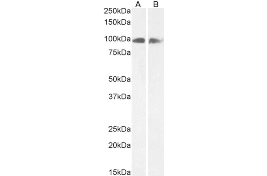
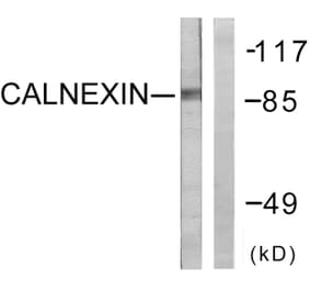
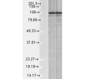
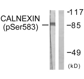
Figure 1: Western Blot - Anti-Calnexin Antibody (A83665)
Calnexin expression in Human Cerebellum (A) and Rat Brain (B) lysate analyzed by western blot. Cells were lysed in RIPA buffer and 35µg protein was run per lane. Primary incubation was performed with Anti-Calnexin Antibody (A83665) at 0.3µg/ml (A) or 0.5µg/ml (B) and detected by chemiluminescence.
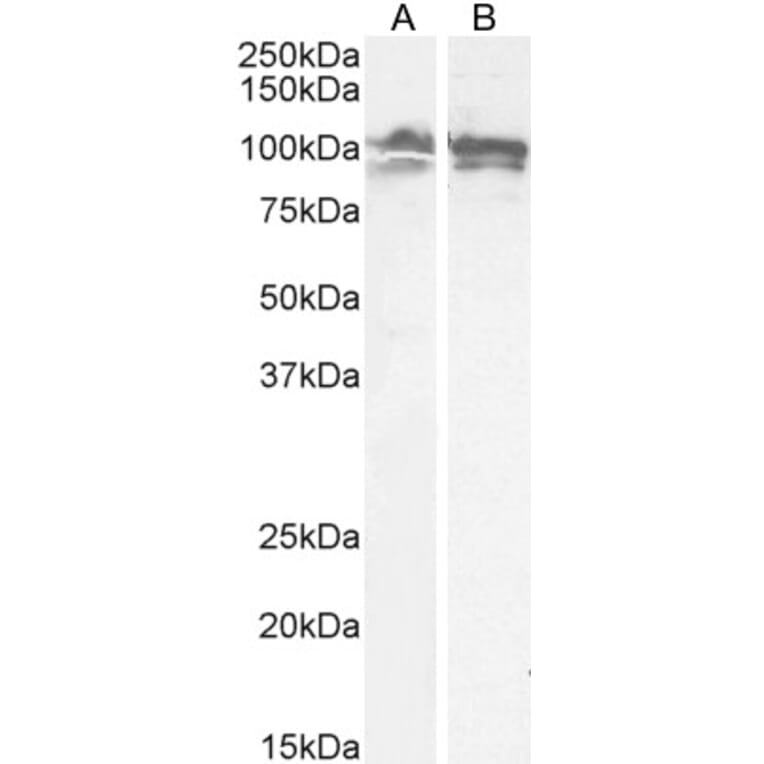
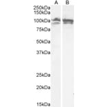
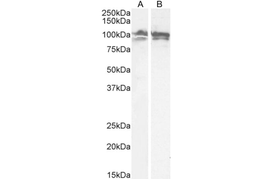
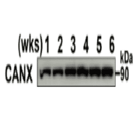
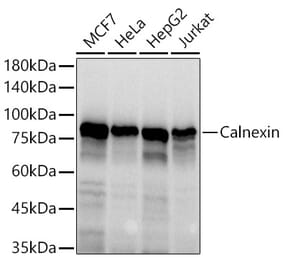
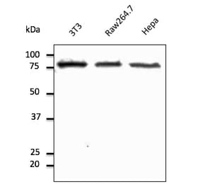
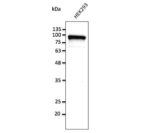
Figure 2: Western Blot - Anti-Calnexin Antibody (A83665)
Calnexin expression in LNCaP (A) and U251 (B) cell lysates analyzed by western blot. Cells were lysed in RIPA buffer and 35µg protein was run per lane. Primary antibody incubation was performed with Anti-Calnexin Antibody (A83665) at 0.5µg/ml and detected by chemiluminescence.
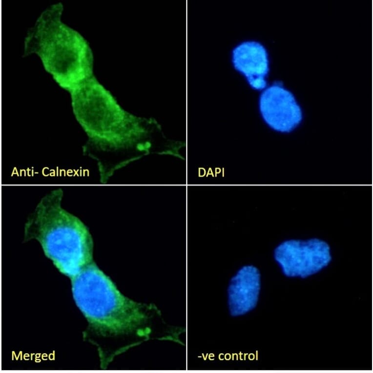
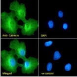
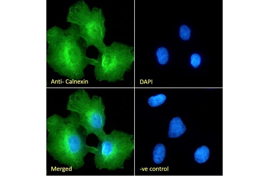
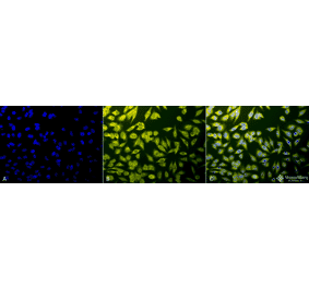
Calnexin expression in U251 cells analyzed by immunofluorescence. Cells were permeabilized with 0.15% Triton. Staining was performed with Anti-Calnexin Antibody (A83665) at 10µg/ml for 1 hour and Alexa Fluor 488 secondary antibody at 2µg/ml. Cytoplasmic and ER staining shown and nuclei were stained with DAPI (blue). Negative control: Goat IgG Isotype Control at 10µg/ml followed by Alexa Fluor 488 secondary antibody at 2µg/ml.
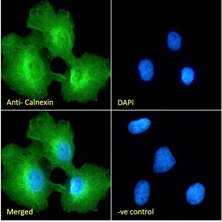
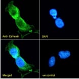
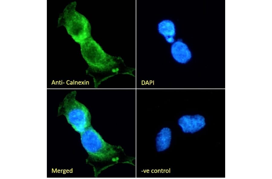
Calnexin expression in LNCaP cells analyzed by immunofluorescence. Cells were permeabilized with 0.15% Triton. Staining was performed with Anti-Calnexin Antibody (A83665) at 10µg/ml for 1 hour and Alexa Fluor 488 secondary antibody at 2µg/ml. ER and plasma membrane staining shown and nuclei were stained with DAPI (blue). Negative control: Goat IgG Isotype Control at 10µg/ml followed by Alexa Fluor 488 secondary antibody at 2µg/ml.
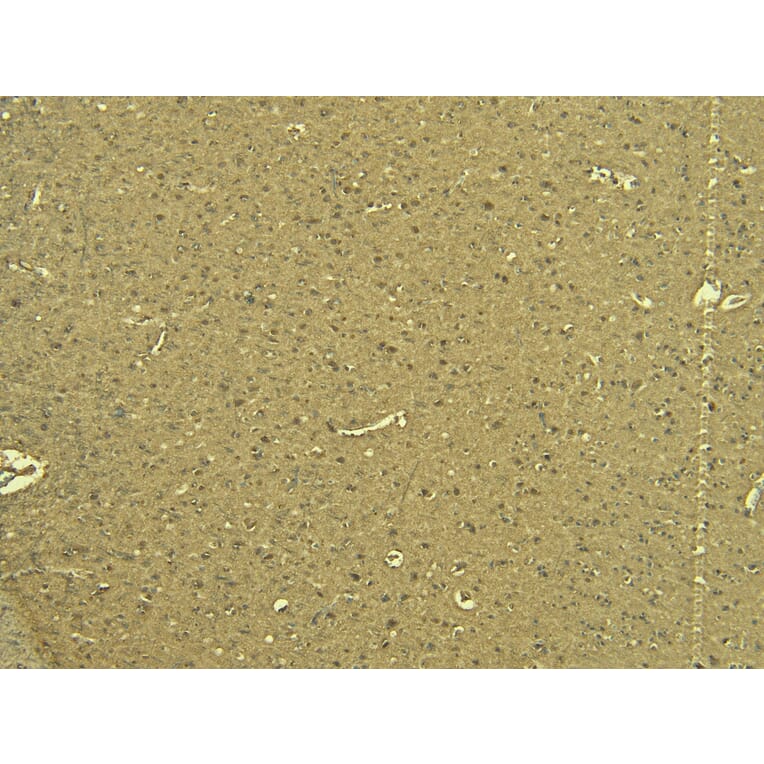
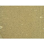
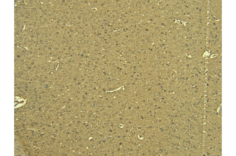
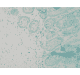
Calnexin expression in Human Cortex analyzed by immunohistochemistry. Tissue was paraffin-embedded, and antigen retrieval was achieved by heating in citrate buffer, pH 6. Staining was performed with Anti-Calnexin Antibody (A83665) at 6µg/ml and revealed with horseradish peroxidase (HRP).
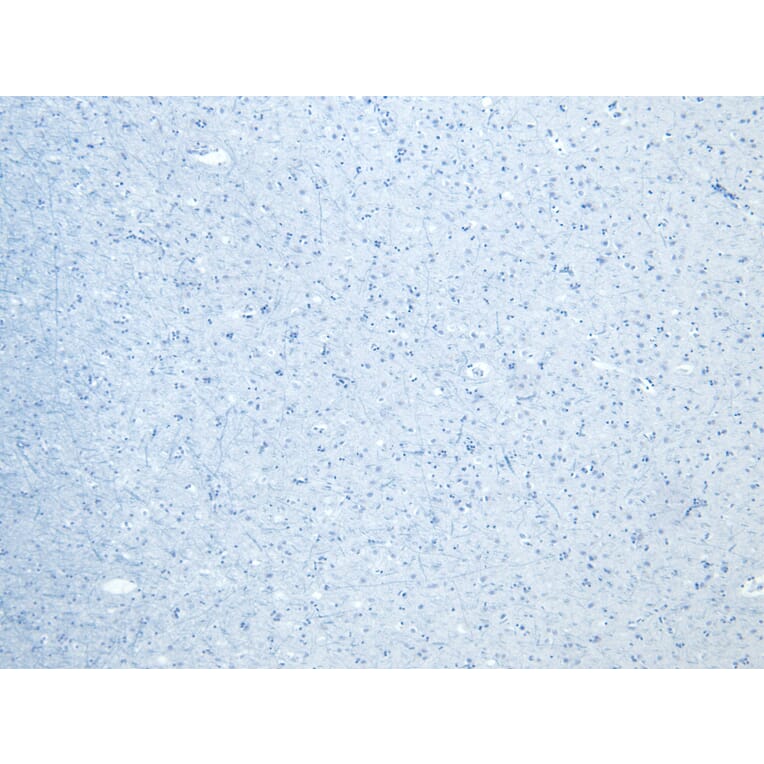
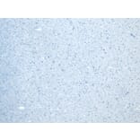
Negative control for Calnexin expression in Human Cortex analyzed by immunohistochemistry. Tissue was paraffin-embedded, and staining procedure was performed in the absence of primary antibody.


![Immunohistochemistry - Anti-Calnexin Antibody [CANX/1541] (A250400) - Antibodies.com](https://cdn.antibodies.com/image/catalog/250/A250400_1.jpg?profile=product_alternative)
![Immunohistochemistry - Anti-Calnexin Antibody [CANX/1541] - BSA and Azide free (A253580) - Antibodies.com](https://cdn.antibodies.com/image/catalog/253/A253580_1.jpg?profile=product_alternative)
![Immunohistochemistry - Anti-Calnexin Antibody [CANX/1543] (A250401) - Antibodies.com](https://cdn.antibodies.com/image/catalog/250/A250401_1.jpg?profile=product_alternative)
![Immunohistochemistry - Anti-Calnexin Antibody [CANX/1543] - BSA and Azide free (A253581) - Antibodies.com](https://cdn.antibodies.com/image/catalog/253/A253581_1.jpg?profile=product_alternative)