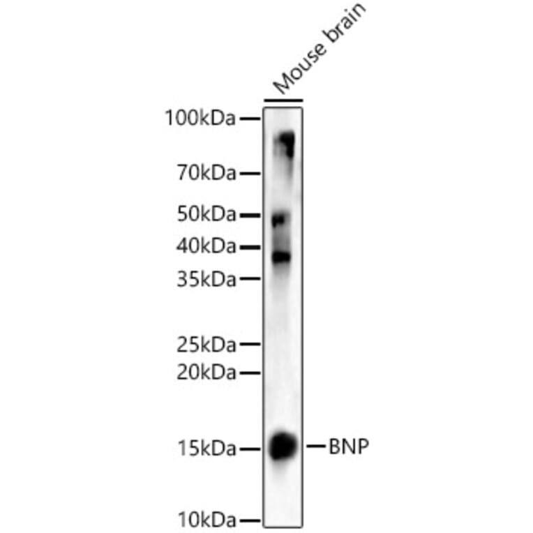
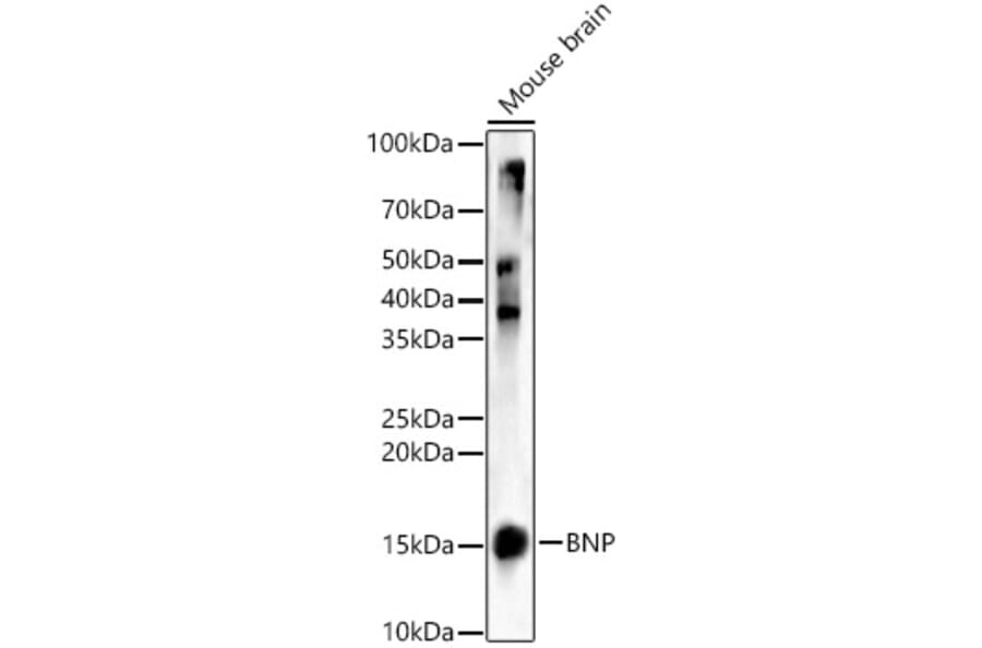
Western blot analysis of Mouse brain, using Anti-BNP Antibody (A11515) at 1:1,000 dilution. The secondary antibody was Goat Anti-Rabbit IgG H&L Antibody (HRP) at 1:10,000 dilution. Lysates/proteins were present at 25µg per lane. The blocking buffer used was 3% non-fat dry milk in TBST. Detection was with a ECL Basic Kit. Exposure time: 30s.
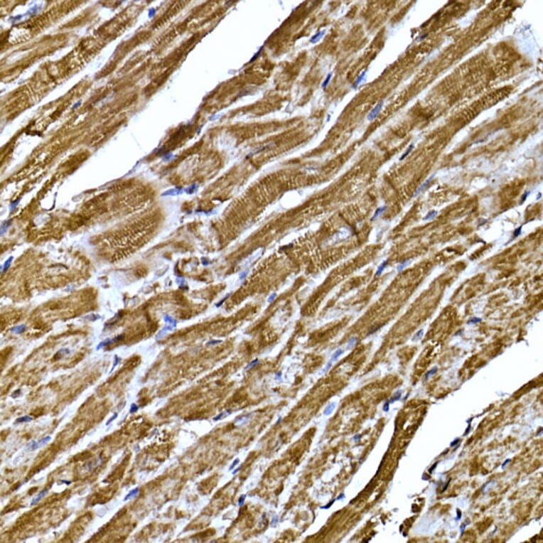
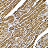
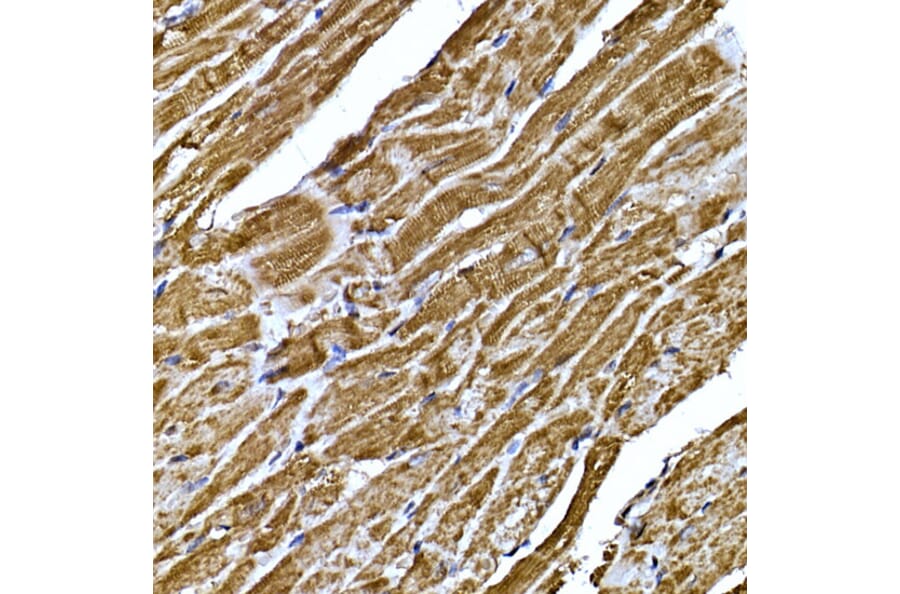
Immunohistochemistry analysis of paraffin-embedded mouse heart using Anti-BNP Antibody (A11515) at a dilution of 1:200 (40x lens). Perform high pressure antigen retrieval with 10 mM citrate buffer pH 6.0 before commencing with IHC staining protocol.
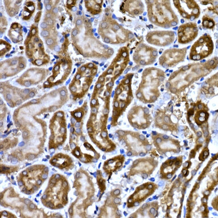
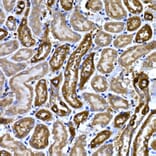
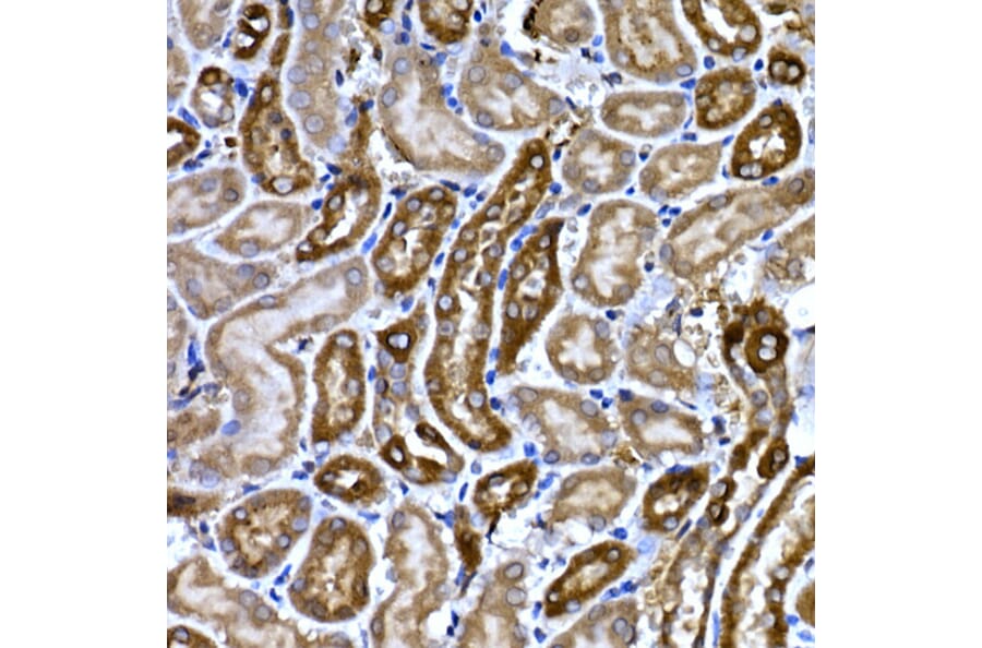
Immunohistochemistry analysis of paraffin-embedded mouse kidney using Anti-BNP Antibody (A11515) at a dilution of 1:200 (40x lens). Perform high pressure antigen retrieval with 10 mM citrate buffer pH 6.0 before commencing with IHC staining protocol.
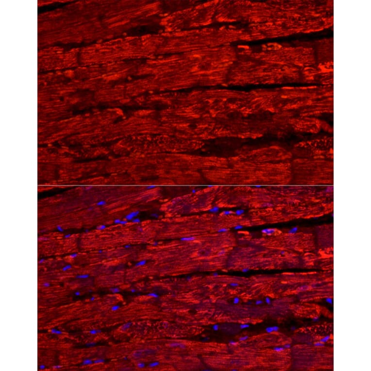
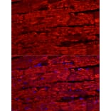
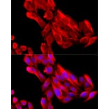
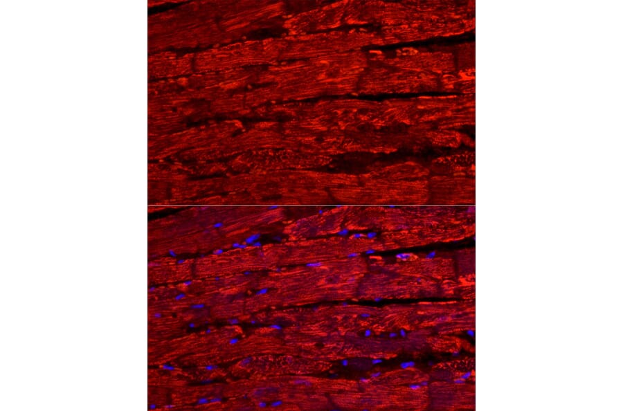
Immunofluorescence analysis of rat heart cells using Anti-BNP Antibody (A11515) at a dilution of 1:200 (40x lens). DAPI was used to stain the cell nuclei (blue). Perform high pressure antigen retrieval with 10 mM citrate buffer pH 6.0 before commencing with IF staining protocol.
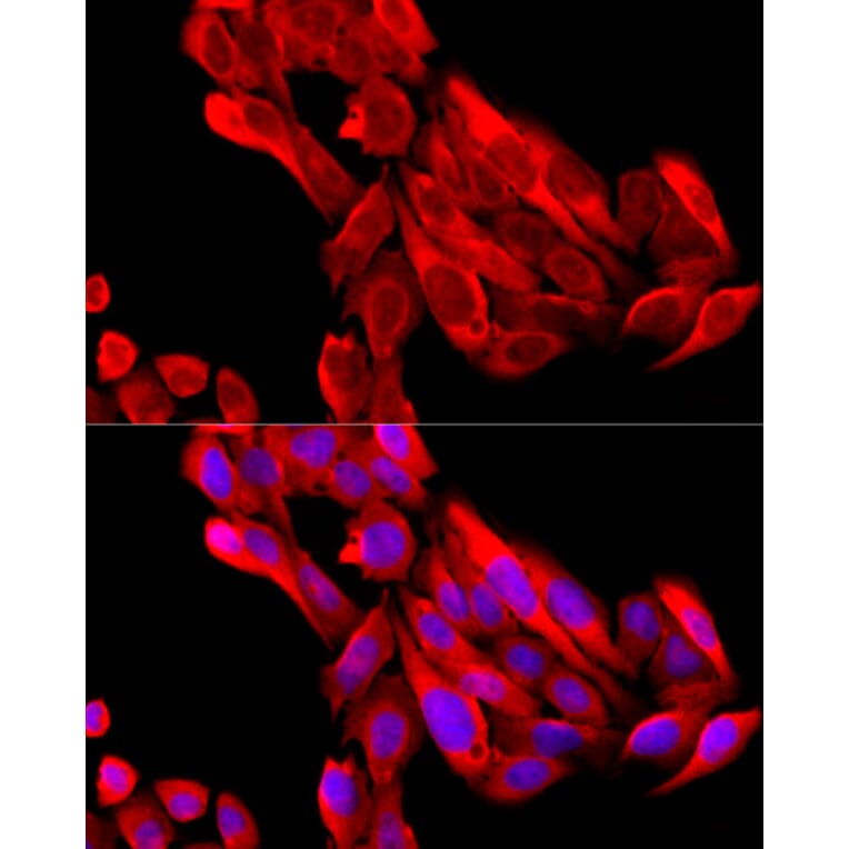
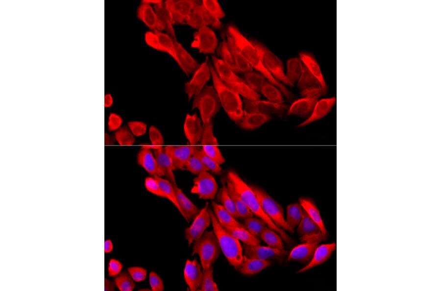
Immunofluorescence analysis of U2OS cells using Anti-BNP Antibody (A11515) at a dilution of 1:200 (40x lens). DAPI was used to stain the cell nuclei (blue).


![Immunohistochemistry - Anti-BNP Antibody [NPPB/4493] - BSA and Azide free (A278316) - Antibodies.com](https://cdn.antibodies.com/image/catalog/278/A278316_1.jpg?profile=product_alternative)
![Immunohistochemistry - Anti-BNP Antibody [NPPB/4493] (A277728) - Antibodies.com](https://cdn.antibodies.com/image/catalog/277/A277728_1.jpg?profile=product_alternative)
