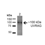
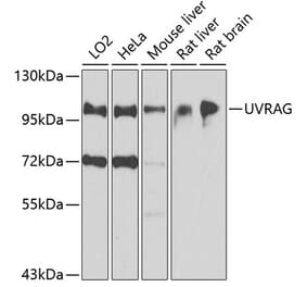
Western blot analysis of rat liver showing detection of ~100kDa UVRAG protein using Anti-UVRAG Antibody (A305065) at 1:200 for 1 hour at room temperature. Lane 1: MW Ladder. Lane 2: rat liver (20 µg). Load: 20 µg. Block: 5% Skim Milk for 1 hour at room temperature. The secondary antibody used was Goat Anti-Rabbit IgG: HRP at 1:2000 for 1 hour at room temperature. Color Development: ECL solution for 6 min at room temperature. Predicted/Observed Size: ~100kDa.
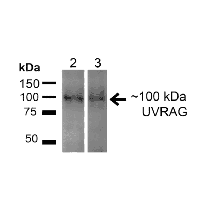
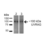
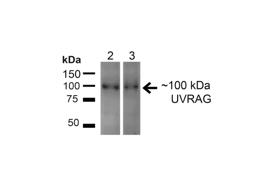
Western blot analysis of human HeLa and HEK293T cell lysates showing detection of ~100kDa UVRAG protein using Anti-UVRAG Antibody (A305065) at 1:200 for 1 hour at room temperature. Lane 1: MW Ladder. Lane 2: human HeLa (20 µg). Lane 3: human 293T (20 µg). Load: 20 µg. Block: 5% Skim Milk for 1 hour at room temperature. The secondary antibody used was Goat Anti-Rabbit IgG: HRP at 1:2000 for 1 hour at room temperature. Color Development: ECL solution for 6 min at room temperature. Predicted/Observed Size: ~100kDa.
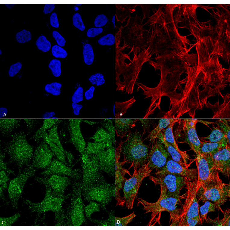
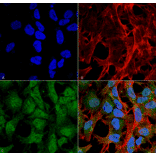
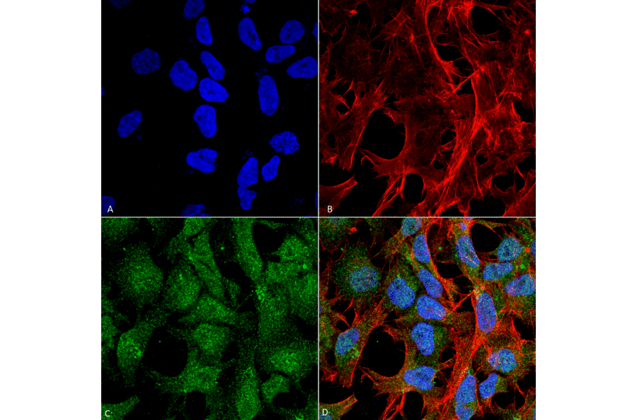
Immunocytochemistry/Immunofluorescence analysis of human neuroblastoma cell line (SK-N-BE, fixed in 4% formaldehyde for 15 min at room temperature, using Anti-UVRAG Antibody (A305065), at 1:100 for 60 minutes at room temperature. The secondary antibody used was Goat Anti-Rabbit ATTO 488 at 1:100 for 60 minutes at room temperature. Counterstain: Phalloidin Texas Red F-Actin stain; DAPI (blue) nuclear stain at 1:1000, 1:5,000 for 60min room temperature, 5min room temperature. Localization: Late Endosome, Lysosome, Early Endosome. Magnification: 60X.(A) DAPI (blue) nuclear stain (B) Phalloidin Texas Red F-Actin stain (C) UVRAG Antibody (D) Composite.
Publishing research using Anti-UVRAG Antibody (A305065)? Please let us know so that we can list the citation on this page.
Alternative products to Anti-UVRAG Antibody (A305065)