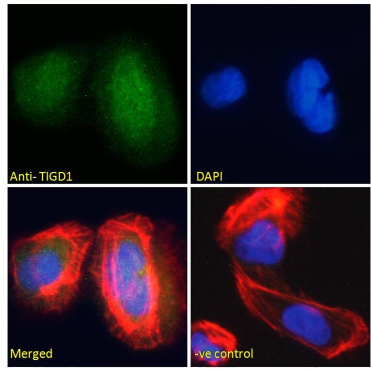
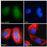
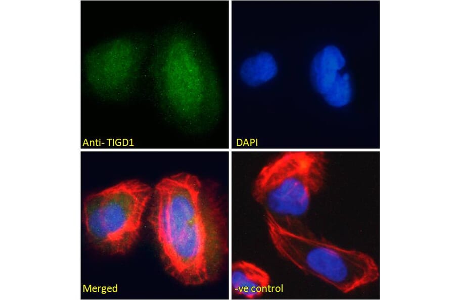
TIGD1 expression in U2OS cells analyzed by immunofluorescence. Cells were permeabilized with 0.15% Triton. Staining was performed with Anti-TIGD1 Antibody (A285939) at 10µg/ml for 1 hour and Alexa Fluor 488 secondary antibody at 2µg/ml. Nuclear staining shown and nuclei were stained with DAPI (blue) while actin filaments were stained with phalloidin (red). Negative control: Goat IgG Isotype Control at 10µg/ml followed by Alexa Fluor 488 secondary antibody at 2µg/ml.
Alternative Produkte zum Anti-TIGD1 Antikörper (A285939)