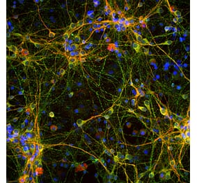
Immunohistochemistry analysis of mouse brain slice. The Primary Antibody used was Anti-Tau (phospho Ser202 + Thr205) Antibody [AH36] (A304919) at 1:500 for Overnight at 4°C. The secondary antibody used was Anti-Rabbit IgG: AlexaFluor 488. Counterstain: DAPI at 1:1,000 for 5 min. CA3 Region of P301SxUBQLN2 Tg mouse. IHC Protocol: 1. Post-fix brains in 4% PFA for 24 hours and put through a 10-30% sucrose gradient. 2. Section by cryostat at 10 uM thickness. 3. Fix in MeOH 15 min. 4. 3 - 10 min wash in PBS 1X. 5. Heat via microwave in 10mM Citrate Buffer, pH 6 for 4 min at power level 20. 6. Cool in solution for 20 min. 7. Wash 2 - 5 minutes in PBS. 8. Permeabilize in 0.5% Triton-X 100 in PBS 10 min. 9. Wash in PBS 10 min. 10. Block for 1 hour in 5% goat serum. 11. Incubate primary Ab (1:500) in blocking solution overnight at 4°C. 12. Wash 3 - 10 min in PBS. 13. Incubate in secondary Ab Rb IgG Alexa-fluor 488. 14. Wash 3 - 10 min in PBS. 15. Incubate in DAPI 1:1,000 for 5 min. 16. Wash 3 - 5 min. 17. Coverslip with Prolong-Gold.
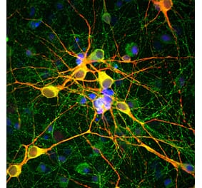
Immunohistochemistry analysis of mouse brain slice. The Primary Antibody used was Anti-Tau (phospho Ser202 + Thr205) Antibody [AH36] (A304919) at 1:500 for Overnight at 4°C. The secondary antibody used was Anti-Rabbit IgG: AlexaFluor 488. Counterstain: DAPI at 1:1,000 for 5 min.(A) Pons of Non-Tg mouse. (B) Pons of P301SxUBQLN2 Tg mouse. (C) Prefrontal cortex of Non-Tg mouse. (D) Prefrontal cortex of P301SxUBQLN2 Tg mouse. IHC Protocol: 1. Post-fix brains in 4% PFA for 24 hours and put through a 10-30% sucrose gradient. 2. Section by cryostat at 10 uM thickness. 3. Fix in MeOH 15 min. 4. 3 - 10 min wash in PBS 1X. 5. Heat via microwave in 10mM Citrate Buffer, pH 6 for 4 min at power level 20. 6. Cool in solution for 20 min. 7. Wash 2 - 5 minutes in PBS. 8. Permeabilize in 0.5% Triton-X 100 in PBS 10 min. 9. Wash in PBS 10 min. 10. Block for 1 hour in 5% goat serum. 11. Incubate primary Ab (at 1:500) in blocking solution overnight at 4°C. 12. Wash 3 - 10 min in PBS. 13. Incubate in secondary Ab Rb IgG Alexa-fluor 488. 14. Wash 3 - 10 min in PBS. 15. Incubate in DAPI 1:1,000 for 5 min. 16. Wash 3 - 5 min. 17. Coverslip with Prolong-Gold.
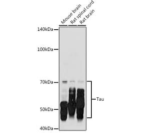
Western Blot - Anti-Tau (phospho Ser202 + Thr205) Antibody [AH36] (A304919)
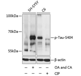
Western blot analysis of human iPSC-derived cortical neurons showing detection of Tau protein using Anti-Tau (phospho Ser202 + Thr205) Antibody [AH36] (A304919) at 1:500 for Overnight. Lane 1: MW ladder. Lane 2: Control (non-disease) line. Lane 2: Ex10+16 tau mutant sample. Lane 3: P301L tau mutant sample. Load: 50ug. pSer202/pThr205 was detected using A304919. Total tau was detected using mouse anti-tau antibody (clone HT7). The bar graph on the right shows quantification of pSer202/pThr205 compared to total tau in each sample.
Dot blot analysis of E. Coli, Baculovirus using Anti-Tau (phospho Ser202 + Thr205) Antibody [AH36] (A304919) at 1:500. The secondary antibody used was Goat anti-rabbit IgG:HRP.
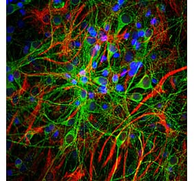
Immunocytochemistry/Immunofluorescence analysis of human iPSC-derived cortical excitatory neurons, using Anti-Tau (phospho Ser202 + Thr205) Antibody [AH36] (A304919), at 1:500 for Overnight. The secondary antibody used was Donkey anti-rabbit: Alexa Fluor 488 at 1:1,000. Counterstain: DAPI. A) iPSC-derived neurons from non-demented control (NDC). B) iPSC-derived neurons from subject with P301L MAPT mutation. Images acquired using an automated Opera Phoenix system. Each field of view is a max projection from 10 planes of 1 μm stacks.
Publishing research using Anti-Tau (phospho Ser202 + Thr205) Antibody [AH36] (A304919)? Please let us know so that we can list the citation on this page.


![Immunohistochemistry - Anti-Tau (phospho Ser202 + Thr205) Antibody [AH36] (A304919) - Antibodies.com](https://cdn.antibodies.com/image/catalog/304/A304919_1.png?profile=product_top)
![Immunohistochemistry - Anti-Tau (phospho Ser202 + Thr205) Antibody [AH36] (A304919) - Antibodies.com](https://cdn.antibodies.com/image/catalog/304/A304919_2.png?profile=product_top)
![Western Blot - Anti-Tau (phospho Ser202 + Thr205) Antibody [AH36] (A304919) - Antibodies.com](https://cdn.antibodies.com/image/catalog/304/A304919_3.png?profile=product_top)
![Dot Blot - Anti-Tau (phospho Ser202 + Thr205) Antibody [AH36] (A304919) - Antibodies.com](https://cdn.antibodies.com/image/catalog/304/A304919_4.png?profile=product_top)
![Immunocytochemistry/Immunofluorescence - Anti-Tau (phospho Ser202 + Thr205) Antibody [AH36] (A304919) - Antibodies.com](https://cdn.antibodies.com/image/catalog/304/A304919_5.png?profile=product_top)
![Immunohistochemistry - Anti-Tau (phospho Ser202 + Thr205) Antibody [AH36] (A304919) - Antibodies.com](https://cdn.antibodies.com/image/catalog/304/A304919_1.png?profile=product_top_thumb)
![Immunohistochemistry - Anti-Tau (phospho Ser202 + Thr205) Antibody [AH36] (A304919) - Antibodies.com](https://cdn.antibodies.com/image/catalog/304/A304919_2.png?profile=product_top_thumb)
![Western Blot - Anti-Tau (phospho Ser202 + Thr205) Antibody [AH36] (A304919) - Antibodies.com](https://cdn.antibodies.com/image/catalog/304/A304919_3.png?profile=product_top_thumb)
![Dot Blot - Anti-Tau (phospho Ser202 + Thr205) Antibody [AH36] (A304919) - Antibodies.com](https://cdn.antibodies.com/image/catalog/304/A304919_4.png?profile=product_top_thumb)
![Immunocytochemistry/Immunofluorescence - Anti-Tau (phospho Ser202 + Thr205) Antibody [AH36] (A304919) - Antibodies.com](https://cdn.antibodies.com/image/catalog/304/A304919_5.png?profile=product_top_thumb)
![Immunohistochemistry - Anti-Tau (phospho Ser202 + Thr205) Antibody [AH36] (A304919) - Antibodies.com](https://cdn.antibodies.com/image/catalog/304/A304919_1.png?profile=product_image)
![Immunohistochemistry - Anti-Tau (phospho Ser202 + Thr205) Antibody [AH36] (A304919) - Antibodies.com](https://cdn.antibodies.com/image/catalog/304/A304919_2.png?profile=product_image)
![Western Blot - Anti-Tau (phospho Ser202 + Thr205) Antibody [AH36] (A304919) - Antibodies.com](https://cdn.antibodies.com/image/catalog/304/A304919_3.png?profile=product_image)
![Dot Blot - Anti-Tau (phospho Ser202 + Thr205) Antibody [AH36] (A304919) - Antibodies.com](https://cdn.antibodies.com/image/catalog/304/A304919_4.png?profile=product_image)
![Immunocytochemistry/Immunofluorescence - Anti-Tau (phospho Ser202 + Thr205) Antibody [AH36] (A304919) - Antibodies.com](https://cdn.antibodies.com/image/catalog/304/A304919_5.png?profile=product_image)
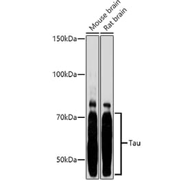
![Western Blot - Anti-Tau Antibody [1D5] (A305068) - Antibodies.com](https://cdn.antibodies.com/image/catalog/305/A305068_1.png?profile=product_alternative)
![Immunocytochemistry/Immunofluorescence - Anti-Tau Antibody [3D4] (A305069) - Antibodies.com](https://cdn.antibodies.com/image/catalog/305/A305069_1.png?profile=product_alternative)
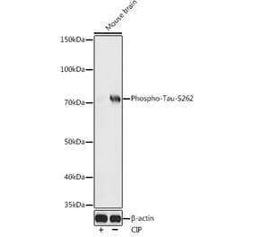
![Western Blot - Anti-Tau (phospho Ser396) Antibody [ARC1573] (A309489) - Antibodies.com](https://cdn.antibodies.com/image/catalog/309/A309489_1.jpg?profile=product_alternative)
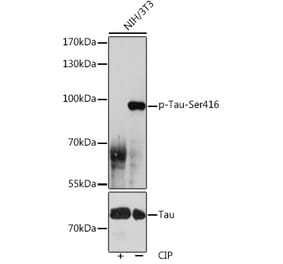
![Western Blot - Anti-Tau (phospho Thr231) Antibody [ARC0021] (A309486) - Antibodies.com](https://cdn.antibodies.com/image/catalog/309/A309486_1.jpg?profile=product_alternative)