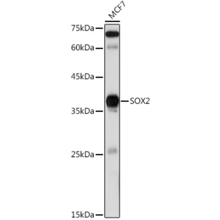
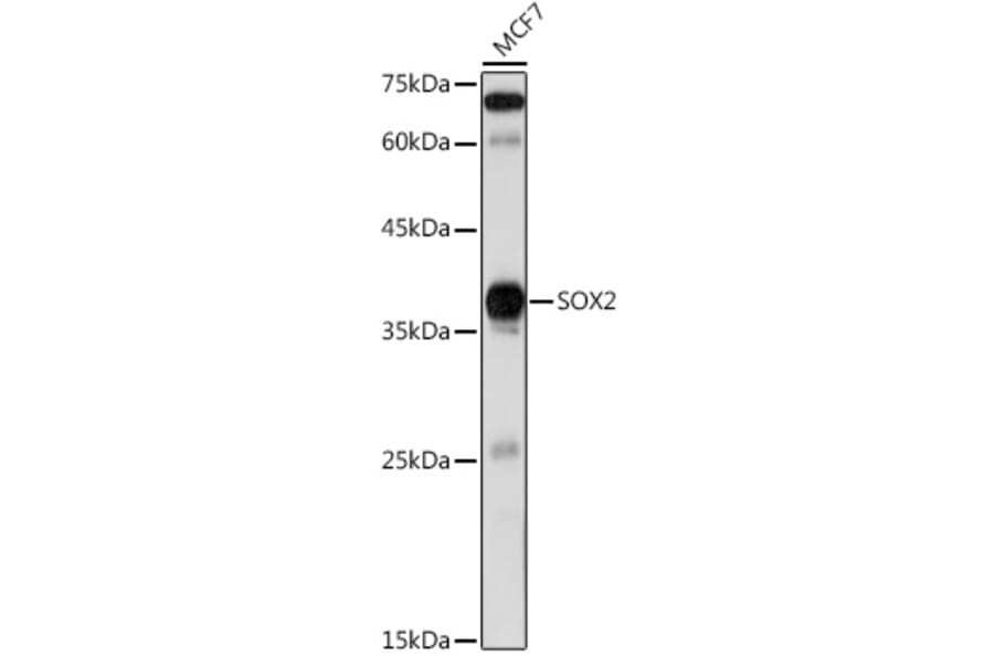
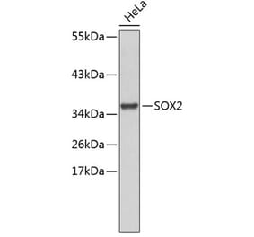
Western blot analysis of extracts of MCF7 cells, using Anti-SOX2 Antibody (A12651) at 1:1,000 dilution. The secondary antibody was Goat Anti-Rabbit IgG H&L Antibody (HRP) at 1:10,000 dilution. Lysates/proteins were present at 25µg per lane. The blocking buffer used was 3% non-fat dry milk in TBST. Detection was with a ECL Basic Kit. Exposure time: 60s.
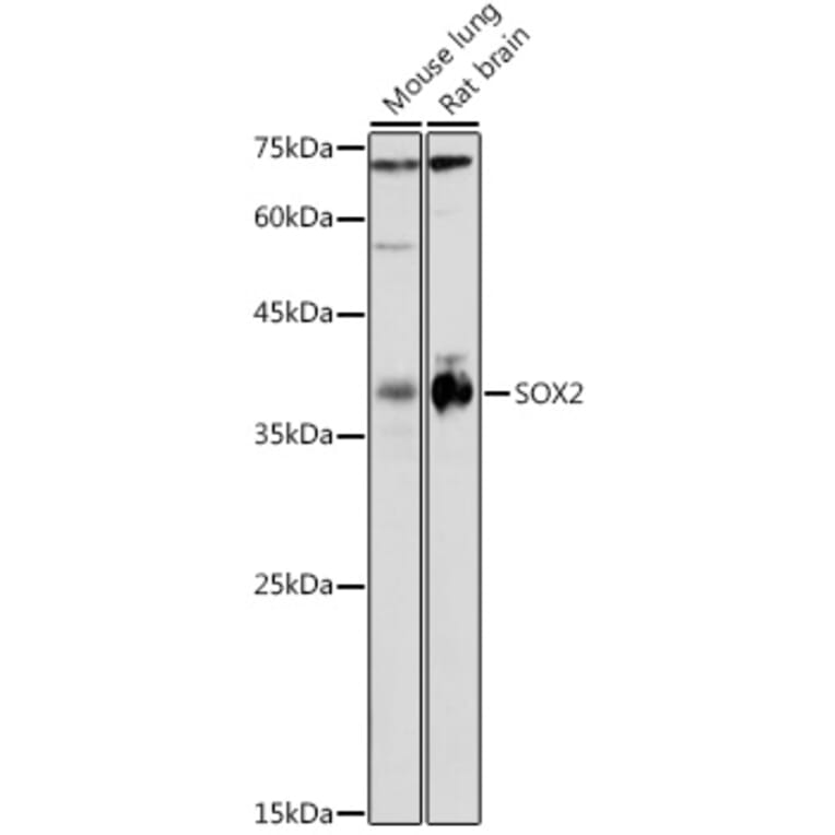
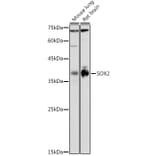
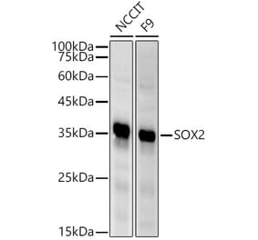
Western blot analysis of extracts of various cell lines, using Anti-SOX2 Antibody (A12651) at 1:1,000 dilution. The secondary antibody was Goat Anti-Rabbit IgG H&L Antibody (HRP) at 1:10,000 dilution. Lysates/proteins were present at 25µg per lane. The blocking buffer used was 3% non-fat dry milk in TBST. Detection was with a ECL Basic Kit. Exposure time: 180s.
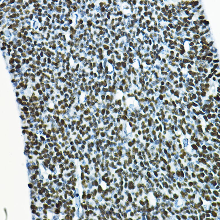
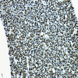
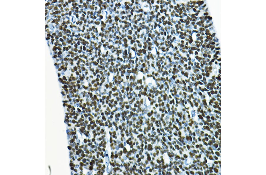
Immunohistochemistry analysis of paraffin-embedded mouse embryos using Anti-SOX2 Antibody (A12651) at a dilution of 1:100 (40x lens). Perform high pressure antigen retrieval with 10 mM citrate buffer pH 6.0 before commencing with IHC staining protocol.
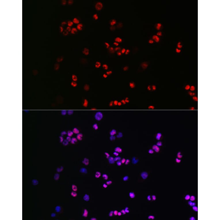
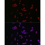
Immunofluorescence analysis of F9 cells using Anti-SOX2 Antibody (A12651) at a dilution of 1:50 (40x lens). DAPI was used to stain the cell nuclei (blue).
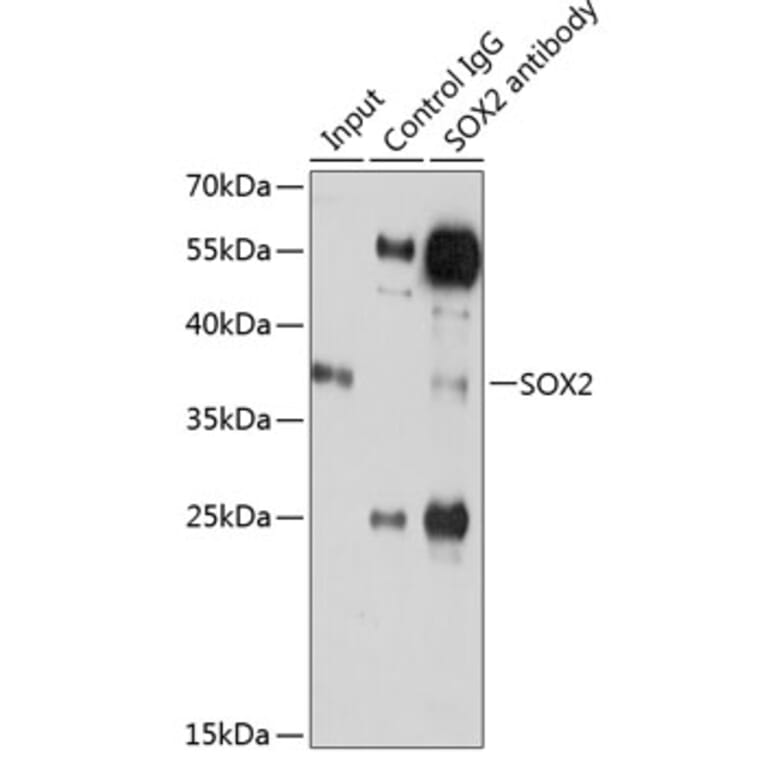
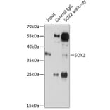
Immunoprecipitation analysis of 200µg extracts of C6 cells using 3µg of Anti-SOX2 Antibody (A12651). This Western blot was performed on the immunoprecipitate using Anti-SOX2 Antibody (A12651) at a dilution of 1:500.


![Immunohistochemistry - Anti-SOX2 Antibody [SOX2/1791] (A250002) - Antibodies.com](https://cdn.antibodies.com/image/catalog/250/A250002_1.jpg?profile=product_alternative)
![Immunohistochemistry - Anti-SOX2 Antibody [SOX2/1791] - BSA and Azide free (A253182) - Antibodies.com](https://cdn.antibodies.com/image/catalog/253/A253182_1.jpg?profile=product_alternative)
![Immunohistochemistry - Anti-SOX2 Antibody [SOX2/1792] (A250003) - Antibodies.com](https://cdn.antibodies.com/image/catalog/250/A250003_1.jpg?profile=product_alternative)
![Immunohistochemistry - Anti-SOX2 Antibody [SOX2/1792] - BSA and Azide free (A253183) - Antibodies.com](https://cdn.antibodies.com/image/catalog/253/A253183_1.jpg?profile=product_alternative)
![Immunohistochemistry - Anti-SOX2 Antibody [SOX2/3169R] - BSA and Azide free (A253185) - Antibodies.com](https://cdn.antibodies.com/image/catalog/253/A253186_1.jpg?profile=product_alternative)

![Immunohistochemistry - Anti-SOX2 Antibody [SOX2/3811R] - BSA and Azide free (A253187) - Antibodies.com](https://cdn.antibodies.com/image/catalog/253/A253187_1.jpg?profile=product_alternative)
![Immunohistochemistry - Anti-SOX2 Antibody [SOX2/3811R] (A250007) - Antibodies.com](https://cdn.antibodies.com/image/catalog/250/A250007_1.jpg?profile=product_alternative)
![SDS-PAGE - Anti-SOX2 Antibody [rSOX2/1792] (A250005) - Antibodies.com](https://cdn.antibodies.com/image/catalog/250/A250005_1.jpg?profile=product_alternative)
![SDS-PAGE - Anti-SOX2 Antibody [rSOX2/1792] - BSA and Azide free (A253185) - Antibodies.com](https://cdn.antibodies.com/image/catalog/253/A253185_1.jpg?profile=product_alternative)