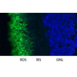
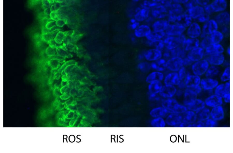
High magnification confocal image of pig retinal section stained with Anti-Rhodopsin Antibody (green). Rhodopsin is most abundant in the rod outer segments (ROS) of retina, clearly localized in rod membranes. The rod inner segments (RIS) and rod nuclei in the outer nuclear layer (ONL) are also seen in this image. Nuclear DNA was stained with DAPI (blue).
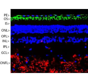
Immunofluorescent analysis of mouse retina section stained with Anti-Rhodopsin Antibody [B630] (A85373), at a dilution of 1:2,000, in green, and co-stained with Anti-Fox3 Antibody (A85403), at a dilution of 1:5,000 in red. The nuclear DNA is visualised in blue using Hoechst staining. The Anti-Rhodopsin Antibody [B630] (A85373) reveals the rod cell membranes located in the photoreceptor outer segments (OS) layer of the retina. The Anti-Fox3 Antibody (A85403) selectively stains the nuclei and cytoplasm of neuronal cells in the ganglion cell layer (GCL), but does not stain most neurons in the layers between.
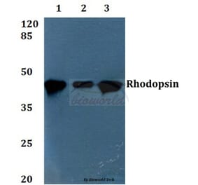
Figure 3: Western Blot - Anti-Rhodopsin Antibody [B630] (A85373)
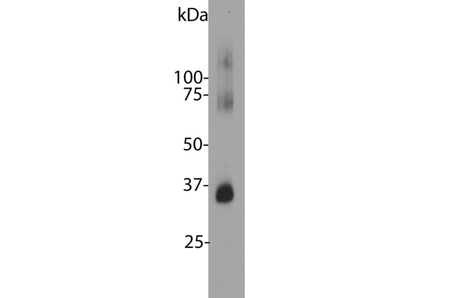
Western blot analysis of retina lysates from different species using Anti-Rhodopsin Antibody [B630] (A85373), at a dilution of 1:5,000, in green. The lanes contain samples of: [1] Protein standards, in red, [2] rat [3] mouse and [4] cow retina lysates. The strong band at 35 kDa corresponds to rhodopsin protein. Bands around 70 kDa and 140 kDa result from the known tendency of rhodopsin to aggregate on SDS-PAGE gels.
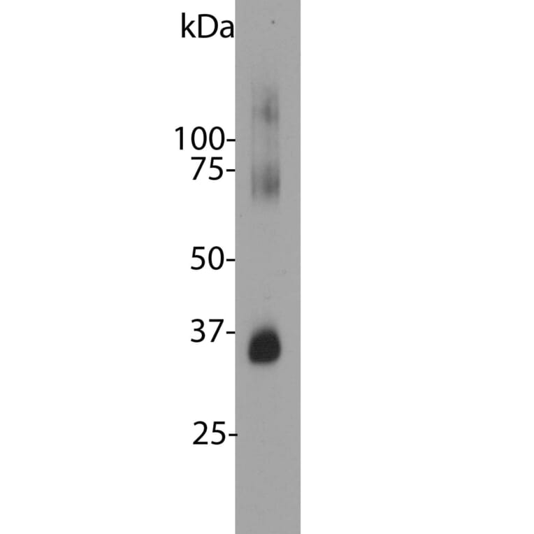
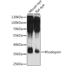
Figure 4: Western Blot - Anti-Rhodopsin Antibody [B630] (A85373)
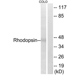
Blot of bovine retinal extracts probed with Anti-Rhodopsin Antibody. The antibody stains a band corresponding to retinal rhodopsin at about 35kDa. Bands about 70 kDa and 140 kDa are aggregated forms of rhodopsin. Note, due to the highly hydrophobic nature of rhodopsin, it is important not to boil a sample containing it in SDS-PAGE sample buffer, as this will result in more extensive aggregation of the rhodopsin protein.
Alternative Produkte zum Anti-Rhodopsin Antikörper [B630] (A85373)


![Immunofluorescence - Anti-Rhodopsin Antibody [B630] (A85373) - Antibodies.com](https://cdn.antibodies.com/image/catalog/85/A85373_3.jpg?profile=product_top)
![Western Blot - Anti-Rhodopsin Antibody [B630] (A85373) - Antibodies.com](https://cdn.antibodies.com/image/catalog/85/A85373_4.jpg?profile=product_top)
![Immunofluorescence - Anti-Rhodopsin Antibody [B630] (A85373) - Antibodies.com](https://cdn.antibodies.com/image/catalog/85/A85373_3.jpg?profile=product_top_thumb)
![Western Blot - Anti-Rhodopsin Antibody [B630] (A85373) - Antibodies.com](https://cdn.antibodies.com/image/catalog/85/A85373_4.jpg?profile=product_top_thumb)
![Immunofluorescence - Anti-Rhodopsin Antibody [B630] (A85373) - Antibodies.com](https://cdn.antibodies.com/image/catalog/85/A85373_3.jpg?profile=product_image)
![Western Blot - Anti-Rhodopsin Antibody [B630] (A85373) - Antibodies.com](https://cdn.antibodies.com/image/catalog/85/A85373_4.jpg?profile=product_image)
![Western Blot - Anti-Rhodopsin Antibody [4D2] (A305179) - Antibodies.com](https://cdn.antibodies.com/image/catalog/305/A305179_1.png?profile=product_alternative)
![Western Blot - Anti-Rhodopsin Antibody [1D4] (A305239) - Antibodies.com](https://cdn.antibodies.com/image/catalog/305/A305239_1.png?profile=product_alternative)

![Western Blot - Anti-Rhodopsin Antibody [ARC1818] (A305833) - Antibodies.com](https://cdn.antibodies.com/image/catalog/305/A305833_1.jpg?profile=product_alternative)