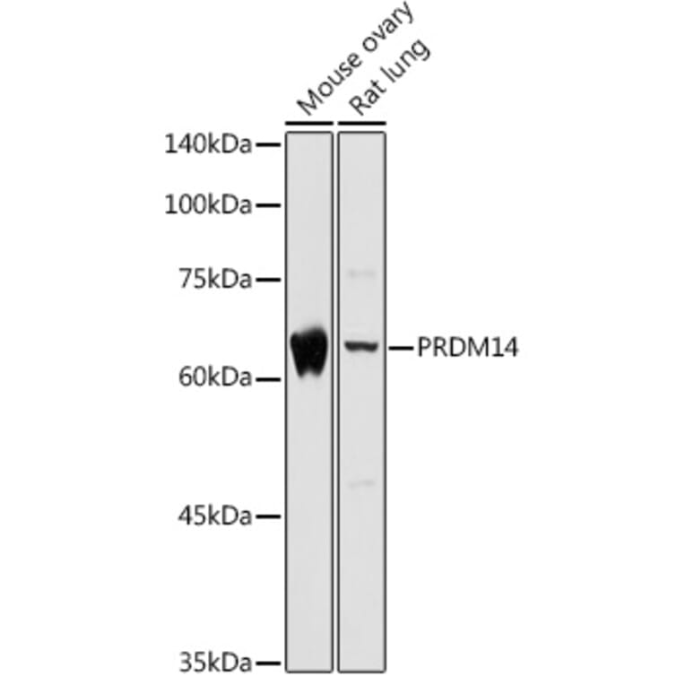
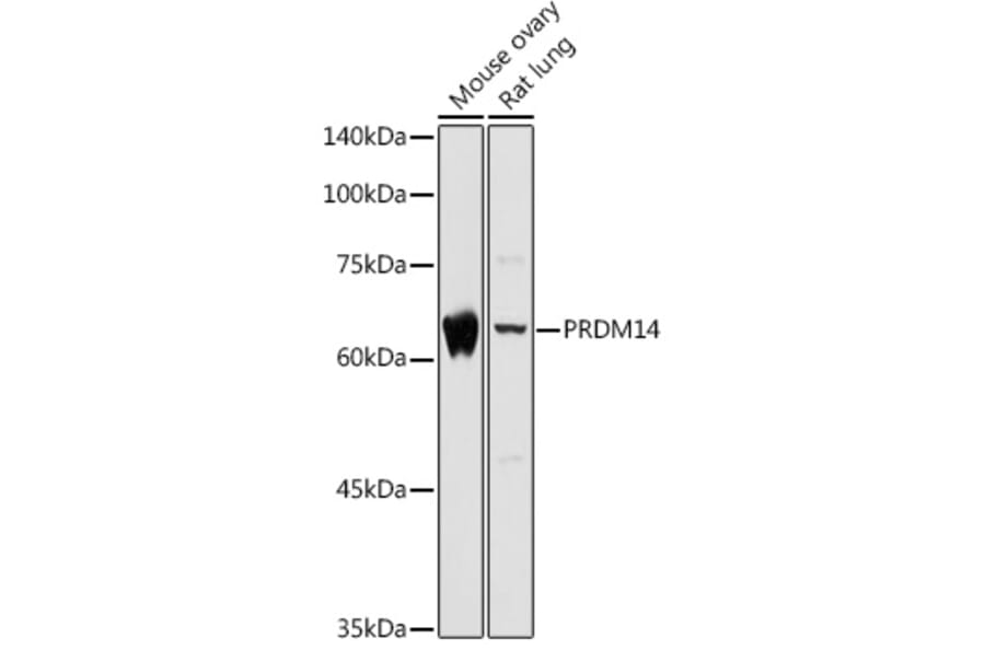
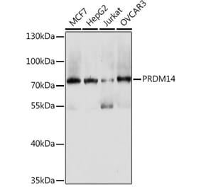
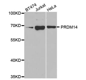
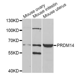
Figure 1: Western Blot - Anti-PRDM14 Antibody (A14825)
Western blot analysis of extracts of various cell lines, using Anti-PRDM14 Antibody (A14825) at 1:1,000 dilution. The secondary antibody was Goat Anti-Rabbit IgG H&L Antibody (HRP) at 1:10,000 dilution. Lysates/proteins were present at 25µg per lane. The blocking buffer used was 3% non-fat dry milk in TBST. Detection was with a ECL Basic Kit. Exposure time: 5s.
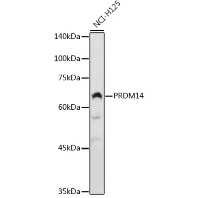
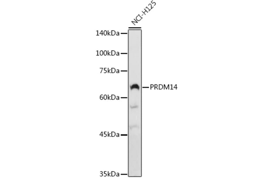
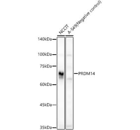
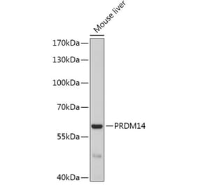
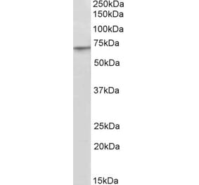
Figure 2: Western Blot - Anti-PRDM14 Antibody (A14825)
Western blot analysis of extracts of NCI-H125 cells, using Anti-PRDM14 Antibody (A14825) at 1:1,000 dilution. The secondary antibody was Goat Anti-Rabbit IgG H&L Antibody (HRP) at 1:10,000 dilution. Lysates/proteins were present at 25µg per lane. The blocking buffer used was 3% non-fat dry milk in TBST. Detection was with a ECL Basic Kit. Exposure time: 90s.
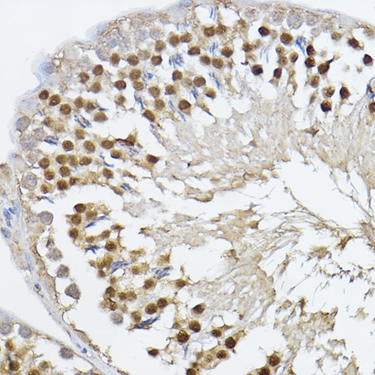
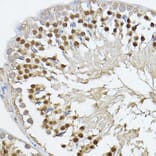
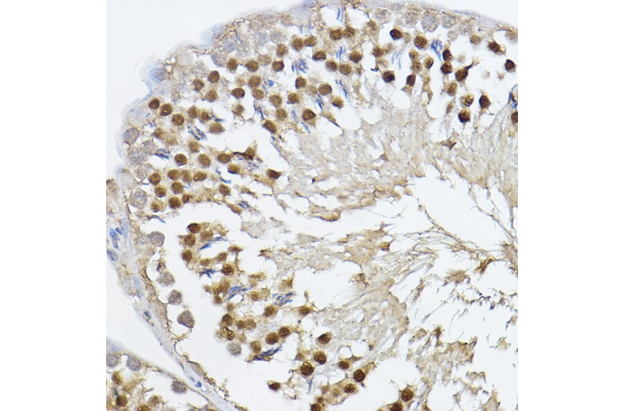
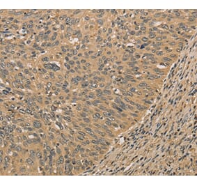
Immunohistochemistry analysis of paraffin-embedded rat testis using Anti-PRDM14 Antibody (A14825) at a dilution of 1:100 (40x lens). Perform high pressure antigen retrieval with 10 mM citrate buffer pH 6.0 before commencing with IHC staining protocol.
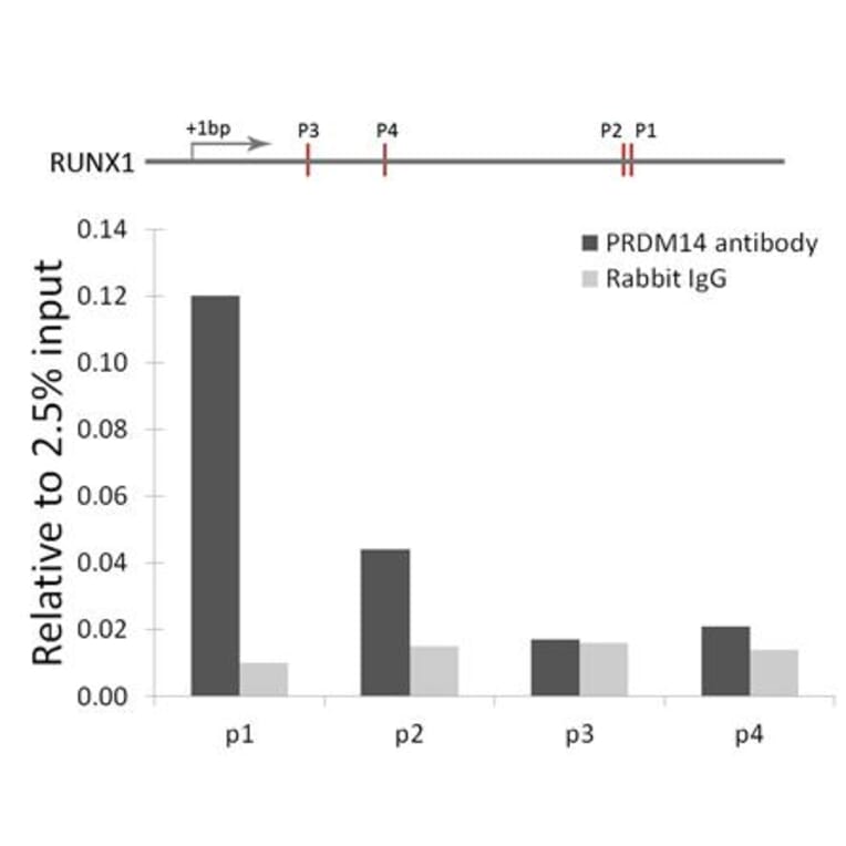
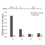
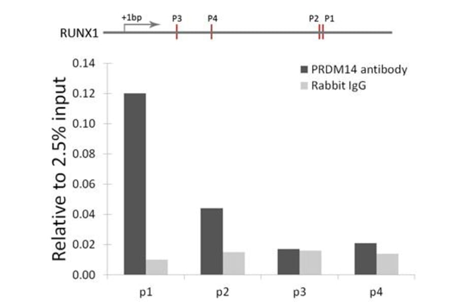
Chromatin immunoprecipitation (ChIP) analysis of RUNX1 gene from 293 cell line, using Anti-PRDM14 Antibody (A14825) and Rabbit IgG. P1, P2, P3 and P4 were four probes located on RUNX1 gene as the schematic diagram illustrated. The amount of immunoprecipitated DNA was checked by quantitative PCR. Histogram was constructed by the ratios of the immunoprecipitated DNA to the input.
Alternative Produkte zum Anti-PRDM14 Antikörper (A14825)