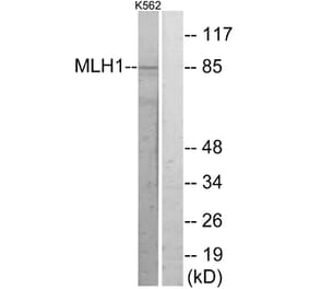
Western Blot - Anti-MLH1 Antibody [ARC53543] (A308155)
Western blot analysis of extracts of various cell lines, using Anti-MLH1 Antibody [ARC53543] (A308155) at 1:1,000 dilution. The secondary antibody was Goat Anti-Rabbit IgG H&L Antibody (HRP) at 1:10,000 dilution. Lysates/proteins were present at 25µg per lane. The blocking buffer used was 3% non-fat dry milk in TBST. Detection was with a ECL Basic Kit. Exposure time: 3s.
Western Blot - Anti-MLH1 Antibody [ARC53543] (A308155)
Western blot analysis of extracts of mouse brain, using Anti-MLH1 Antibody [ARC53543] (A308155) at 1:60,000 dilution. The secondary antibody was Goat Anti-Rabbit IgG H&L Antibody (HRP) at 1:10,000 dilution. Lysates/proteins were present at 25µg per lane. The blocking buffer used was 3% non-fat dry milk in TBST. Detection was with a ECL Basic Kit. Exposure time: 180s.
Western Blot - Anti-MLH1 Antibody [ARC53543] (A308155)
Western blot analysis of extracts from normal (control) and MLH1 knockout (KO) HeLa cells, using Anti-MLH1 Antibody [ARC53543] (A308155) at 1:60,000 dilution. The secondary antibody was Goat Anti-Rabbit IgG H&L Antibody (HRP) at 1:10,000 dilution. Lysates/proteins were present at 25µg per lane. The blocking buffer used was 3% non-fat dry milk in TBST. Detection was with a ECL Basic Kit. Exposure time: 180s.
Immunohistochemistry analysis of paraffin-embedded human colon cancer loss of MLH1 expression (negative control sample) using Anti-MLH1 Antibody [ARC53543] (A308155) at a dilution of 1:200(40x lens). Perform high pressure antigen retrieval with 10 mM citrate buffer pH 6.0 before commencing with IHC staining protocol.
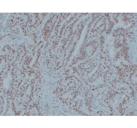
Immunohistochemistry analysis of paraffin-embedded human colon carcinoma tissue using Anti-MLH1 Antibody [ARC53543] (A308155) at a dilution of 1:200(40x lens). Perform high pressure antigen retrieval with 10 mM citrate buffer pH 6.0 before commencing with IHC staining protocol.


![Western Blot - Anti-MLH1 Antibody [ARC53543] (A308155) - Antibodies.com](https://cdn.antibodies.com/image/catalog/308/A308155_1.jpg?profile=product_top)
![Western Blot - Anti-MLH1 Antibody [ARC53543] (A308155) - Antibodies.com](https://cdn.antibodies.com/image/catalog/308/A308155_2.jpg?profile=product_top)
![Western Blot - Anti-MLH1 Antibody [ARC53543] (A308155) - Antibodies.com](https://cdn.antibodies.com/image/catalog/308/A308155_3.jpg?profile=product_top)
![Immunohistochemistry - Anti-MLH1 Antibody [ARC53543] (A308155) - Antibodies.com](https://cdn.antibodies.com/image/catalog/308/A308155_4.jpg?profile=product_top)
![Immunohistochemistry - Anti-MLH1 Antibody [ARC53543] (A308155) - Antibodies.com](https://cdn.antibodies.com/image/catalog/308/A308155_5.jpg?profile=product_top)
![Western Blot - Anti-MLH1 Antibody [ARC53543] (A308155) - Antibodies.com](https://cdn.antibodies.com/image/catalog/308/A308155_1.jpg?profile=product_top_thumb)
![Western Blot - Anti-MLH1 Antibody [ARC53543] (A308155) - Antibodies.com](https://cdn.antibodies.com/image/catalog/308/A308155_2.jpg?profile=product_top_thumb)
![Western Blot - Anti-MLH1 Antibody [ARC53543] (A308155) - Antibodies.com](https://cdn.antibodies.com/image/catalog/308/A308155_3.jpg?profile=product_top_thumb)
![Immunohistochemistry - Anti-MLH1 Antibody [ARC53543] (A308155) - Antibodies.com](https://cdn.antibodies.com/image/catalog/308/A308155_4.jpg?profile=product_top_thumb)
![Immunohistochemistry - Anti-MLH1 Antibody [ARC53543] (A308155) - Antibodies.com](https://cdn.antibodies.com/image/catalog/308/A308155_5.jpg?profile=product_top_thumb)
![Western Blot - Anti-MLH1 Antibody [ARC53543] (A308155) - Antibodies.com](https://cdn.antibodies.com/image/catalog/308/A308155_1.jpg?profile=product_image)
![Western Blot - Anti-MLH1 Antibody [ARC53543] (A308155) - Antibodies.com](https://cdn.antibodies.com/image/catalog/308/A308155_2.jpg?profile=product_image)
![Western Blot - Anti-MLH1 Antibody [ARC53543] (A308155) - Antibodies.com](https://cdn.antibodies.com/image/catalog/308/A308155_3.jpg?profile=product_image)
![Immunohistochemistry - Anti-MLH1 Antibody [ARC53543] (A308155) - Antibodies.com](https://cdn.antibodies.com/image/catalog/308/A308155_4.jpg?profile=product_image)
![Immunohistochemistry - Anti-MLH1 Antibody [ARC53543] (A308155) - Antibodies.com](https://cdn.antibodies.com/image/catalog/308/A308155_5.jpg?profile=product_image)

![Immunohistochemistry - Anti-MLH1 Antibody [MLH1/6467] (A277901) - Antibodies.com](https://cdn.antibodies.com/image/catalog/277/A277901_1.jpg?profile=product_alternative)
![Immunohistochemistry - Anti-MLH1 Antibody [MLH1/6467] - BSA and Azide free (A278489) - Antibodies.com](https://cdn.antibodies.com/image/catalog/278/A278489_1.jpg?profile=product_alternative)
![Immunohistochemistry - Anti-MLH1 Antibody [MLH1/6284R] (A278029) - Antibodies.com](https://cdn.antibodies.com/image/catalog/278/A278029_1.jpg?profile=product_alternative)
![Immunohistochemistry - Anti-MLH1 Antibody [MLH1/6284R] - BSA and Azide free (A278617) - Antibodies.com](https://cdn.antibodies.com/image/catalog/278/A278617_1.jpg?profile=product_alternative)
![Western Blot - Anti-MLH1 Antibody [AMC0417] (A309408) - Antibodies.com](https://cdn.antibodies.com/image/catalog/309/A309408_1.jpg?profile=product_alternative)
![Immunohistochemistry - Anti-MLH1 Antibody [G168-728] - BSA and Azide free (A252531) - Antibodies.com](https://cdn.antibodies.com/image/catalog/252/A252532_1.jpg?profile=product_alternative)
![SDS-PAGE - Anti-MLH1 Antibody [MLH1/6710] (A277711) - Antibodies.com](https://cdn.antibodies.com/image/catalog/277/A277711_1.jpg?profile=product_alternative)
![SDS-PAGE - Anti-MLH1 Antibody [MLH1/6710] - BSA and Azide free (A278299) - Antibodies.com](https://cdn.antibodies.com/image/catalog/278/A278299_1.jpg?profile=product_alternative)