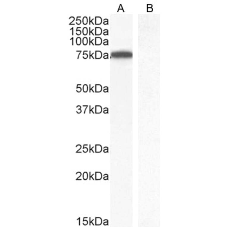
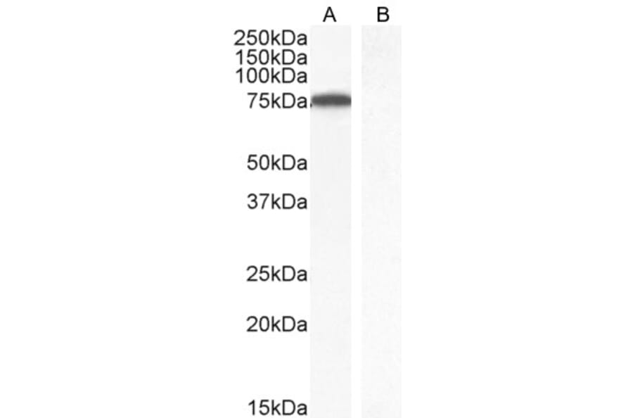
Figure 1: Western Blot - Anti-ITK/EMT Antibody (A83776)
ITK/EMT expression in Jurkat nuclear cell lysate (A) and negative control Human parathyroid gland (B) analyzed by western blot. Cells were lysed in RIPA buffer and 35µg protein was run per lane. Primary antibody incubation was performed with Anti-ITK/EMT Antibody (A83776) at 1µg/ml and detected by chemiluminescence.
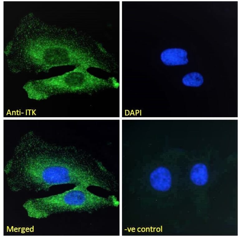
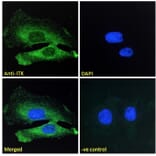
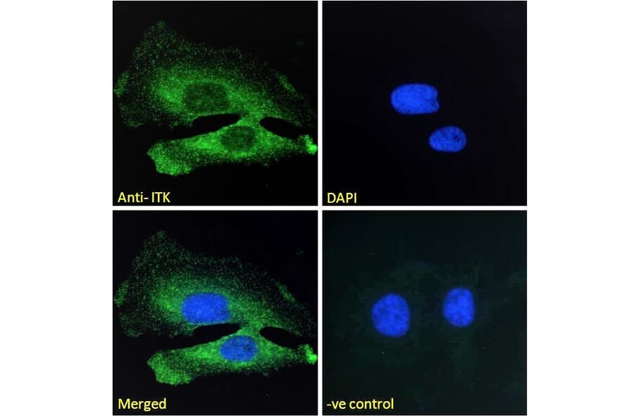
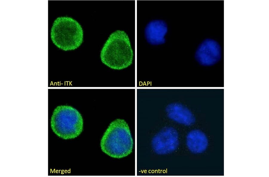
ITK/EMT expression in HeLa cells analyzed by immunofluorescence. Cells were permeabilized with 0.15% Triton. Staining was performed with Anti-ITK/EMT Antibody (A83776) at 10µg/ml for 1 hour and Alexa Fluor 488 secondary antibody at 2µg/ml. Cytoplasmic staining shown and nuclei were stained with DAPI (blue). Negative control: Goat IgG Isotype Control at 10µg/ml followed by Alexa Fluor 488 secondary antibody at 2µg/ml.
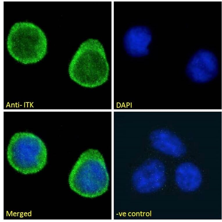
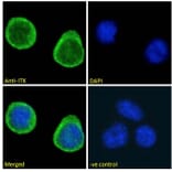
ITK/EMT expression in Jurkat cells analyzed by immunofluorescence. Cells were permeabilized with 0.15% Triton. Staining was performed with Anti-ITK/EMT Antibody (A83776) at 10µg/ml for 1 hour and Alexa Fluor 488 secondary antibody at 2µg/ml. Strong cytoplasmic and weak nuclear staining shown and nuclei were stained with DAPI (blue). Negative control: Goat IgG Isotype Control at 10µg/ml followed by Alexa Fluor 488 secondary antibody at 2µg/ml.