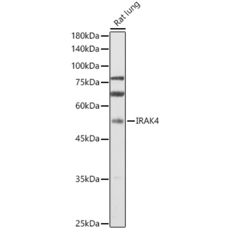
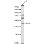
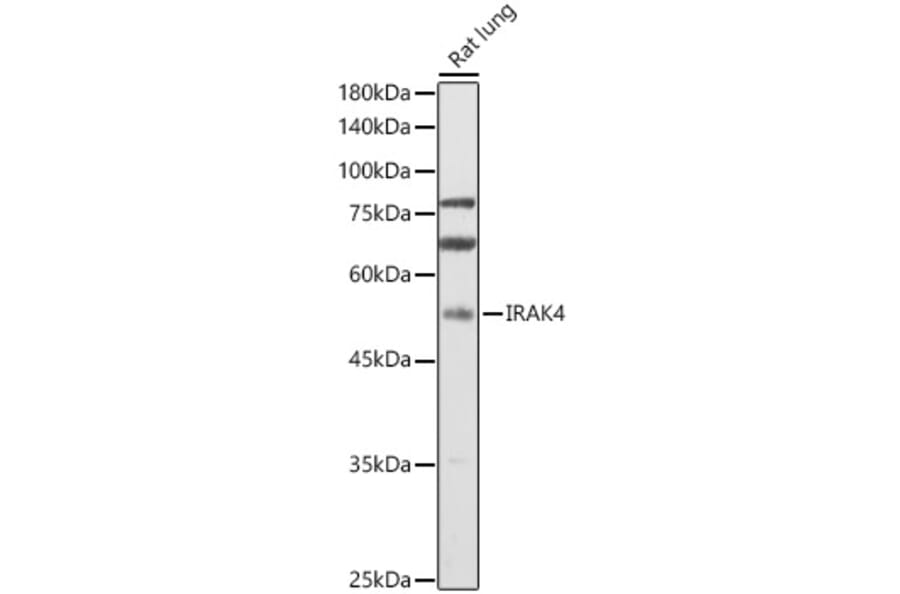
Western blot analysis of extracts of Rat lung, using Anti-IRAK4 Antibody (A15105) at 1:1,000 dilution. The secondary antibody was Goat Anti-Rabbit IgG H&L Antibody (HRP) at 1:10,000 dilution. Lysates/proteins were present at 25µg per lane. The blocking buffer used was 3% non-fat dry milk in TBST. Detection was with a ECL Enhanced Kit (RM00021). Exposure time: 60s.
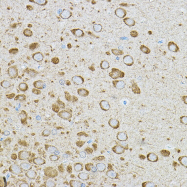
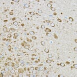
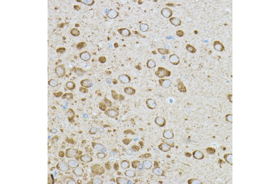
Immunohistochemistry analysis of paraffin-embedded rat brain using Anti-IRAK4 Antibody (A15105) at a dilution of 1:50 (40x lens). Perform high pressure antigen retrieval with 10 mM citrate buffer pH 6.0 before commencing with IHC staining protocol.
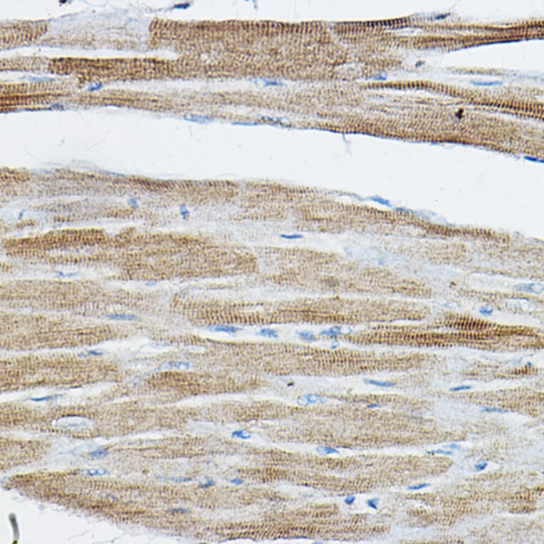
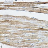
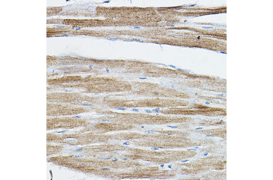
Immunohistochemistry analysis of paraffin-embedded rat heart using Anti-IRAK4 Antibody (A15105) at a dilution of 1:50 (40x lens). Perform high pressure antigen retrieval with 10 mM citrate buffer pH 6.0 before commencing with IHC staining protocol.
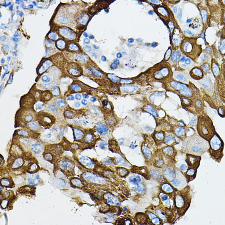
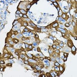
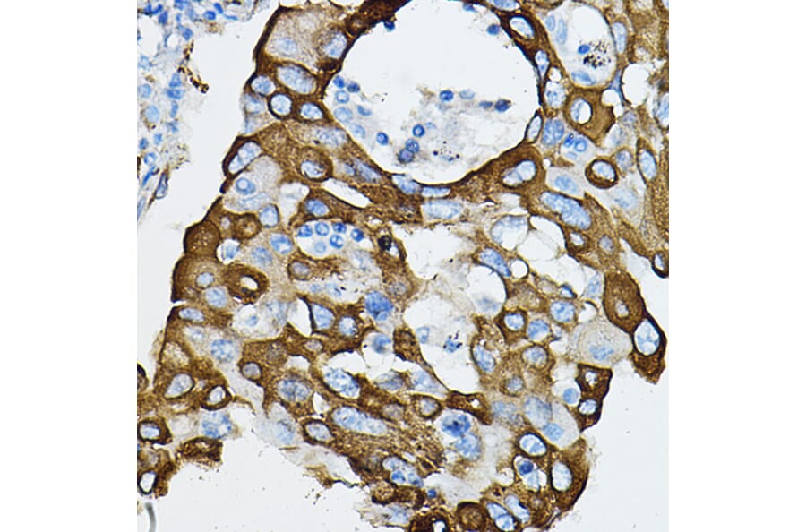
Immunohistochemistry analysis of paraffin-embedded human lung cancer using Anti-IRAK4 Antibody (A15105) at a dilution of 1:50 (40x lens). Perform high pressure antigen retrieval with 10 mM citrate buffer pH 6.0 before commencing with IHC staining protocol.
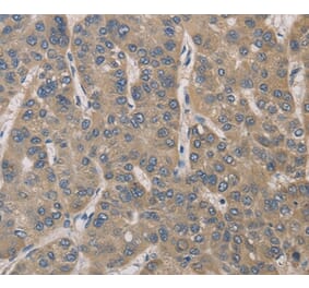
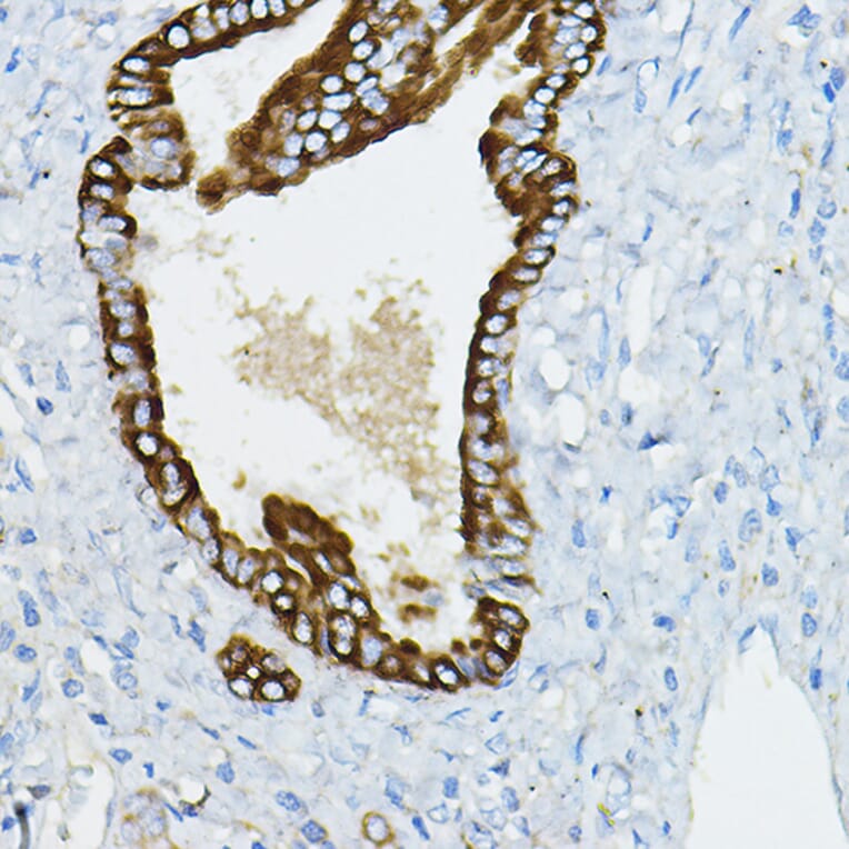
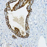
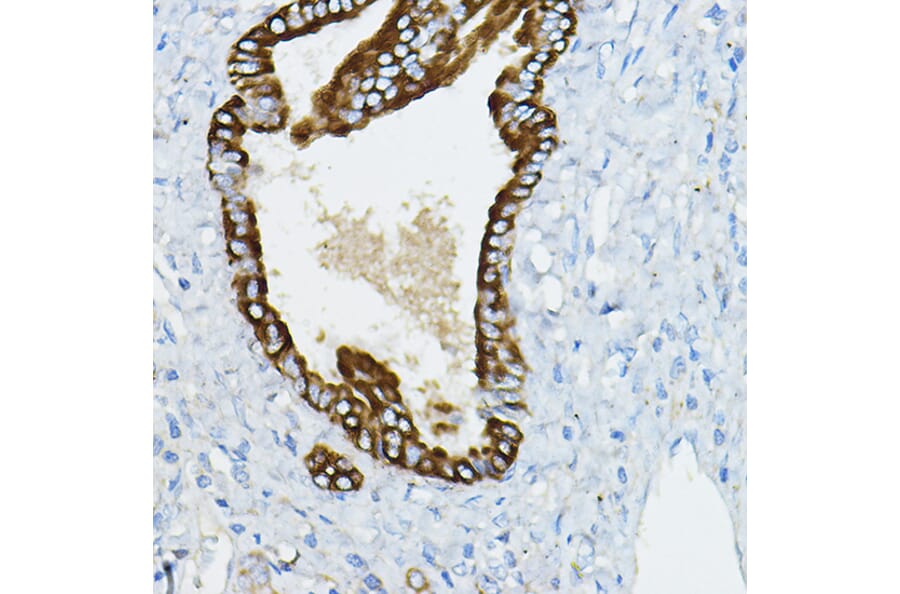
Immunohistochemistry analysis of paraffin-embedded human hepatobiliary duct using Anti-IRAK4 Antibody (A15105) at a dilution of 1:50 (40x lens). Perform high pressure antigen retrieval with 10 mM citrate buffer pH 6.0 before commencing with IHC staining protocol.
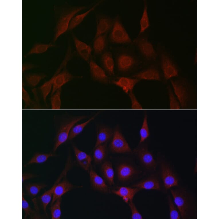
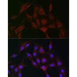
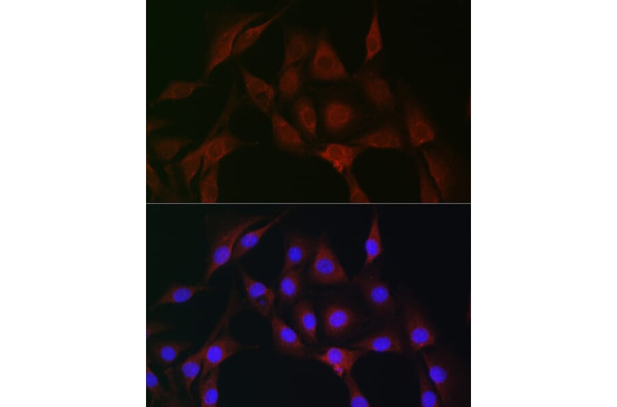
Immunofluorescence analysis of NIH/3T3 cells using Anti-IRAK4 Antibody (A15105) at a dilution of 1:100 (40x lens). DAPI was used to stain the cell nuclei (blue).


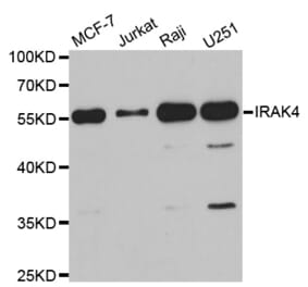
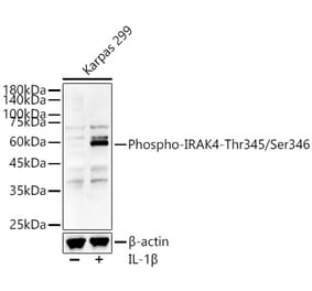
![Western Blot - Anti-IRAK4 (Phospho Ser346) Antibody [ARC64221] (A329540) - Antibodies.com](https://cdn.antibodies.com/image/catalog/329/A329540_1.jpg?profile=product_alternative)
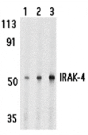
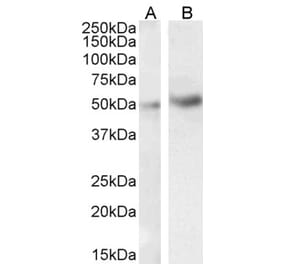
![Western Blot - Anti-IRAK4 Antibody [ARC51289] (A307560) - Antibodies.com](https://cdn.antibodies.com/image/catalog/307/A307560_1.jpg?profile=product_alternative)