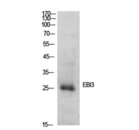
Western Blot - Anti-EBI3 Antibody [ARC2190] (A305621)
Western blot analysis of extracts of MCF7 cells, using Anti-EBI3 Antibody [ARC2190] (A305621) at 1:1,000 dilution. The secondary antibody was Goat Anti-Rabbit IgG H&L Antibody (HRP) at 1:10,000 dilution. Lysates/proteins were present at 25µg per lane. The blocking buffer used was 3% non-fat dry milk in TBST. Detection was with a ECL Basic Kit. Exposure time: 10s.
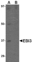
Western Blot - Anti-EBI3 Antibody [ARC2190] (A305621)
Western blot analysis of extracts of various cell lines, using Anti-EBI3 Antibody [ARC2190] (A305621) at 1:1,000 dilution. The secondary antibody was Goat Anti-Rabbit IgG H&L Antibody (HRP) at 1:10,000 dilution. Lysates/proteins were present at 25µg per lane. The blocking buffer used was 3% non-fat dry milk in TBST. Detection was with a ECL Basic Kit. Exposure time: 90s.
Immunofluorescence analysis of rat spleen cells using Anti-EBI3 Antibody [ARC2190] (A305621) at a dilution of 1:100 (40x lens). DAPI was used to stain the cell nuclei (blue).
Immunofluorescence analysis of human spleen cells using Anti-EBI3 Antibody [ARC2190] (A305621) at a dilution of 1:100 (40x lens). DAPI was used to stain the cell nuclei (blue).
Immunofluorescence analysis of mouse spleen cells using Anti-EBI3 Antibody [ARC2190] (A305621) at a dilution of 1:100 (40x lens). DAPI was used to stain the cell nuclei (blue).


![Western Blot - Anti-EBI3 Antibody [ARC2190] (A305621) - Antibodies.com](https://cdn.antibodies.com/image/catalog/305/A305621_1.jpg?profile=product_top)
![Western Blot - Anti-EBI3 Antibody [ARC2190] (A305621) - Antibodies.com](https://cdn.antibodies.com/image/catalog/305/A305621_2.jpg?profile=product_top)
![Immunofluorescence - Anti-EBI3 Antibody [ARC2190] (A305621) - Antibodies.com](https://cdn.antibodies.com/image/catalog/305/A305621_3.jpg?profile=product_top)
![Immunofluorescence - Anti-EBI3 Antibody [ARC2190] (A305621) - Antibodies.com](https://cdn.antibodies.com/image/catalog/305/A305621_4.jpg?profile=product_top)
![Immunofluorescence - Anti-EBI3 Antibody [ARC2190] (A305621) - Antibodies.com](https://cdn.antibodies.com/image/catalog/305/A305621_5.jpg?profile=product_top)
![Western Blot - Anti-EBI3 Antibody [ARC2190] (A305621) - Antibodies.com](https://cdn.antibodies.com/image/catalog/305/A305621_1.jpg?profile=product_top_thumb)
![Western Blot - Anti-EBI3 Antibody [ARC2190] (A305621) - Antibodies.com](https://cdn.antibodies.com/image/catalog/305/A305621_2.jpg?profile=product_top_thumb)
![Immunofluorescence - Anti-EBI3 Antibody [ARC2190] (A305621) - Antibodies.com](https://cdn.antibodies.com/image/catalog/305/A305621_3.jpg?profile=product_top_thumb)
![Immunofluorescence - Anti-EBI3 Antibody [ARC2190] (A305621) - Antibodies.com](https://cdn.antibodies.com/image/catalog/305/A305621_4.jpg?profile=product_top_thumb)
![Immunofluorescence - Anti-EBI3 Antibody [ARC2190] (A305621) - Antibodies.com](https://cdn.antibodies.com/image/catalog/305/A305621_5.jpg?profile=product_top_thumb)
![Western Blot - Anti-EBI3 Antibody [ARC2190] (A305621) - Antibodies.com](https://cdn.antibodies.com/image/catalog/305/A305621_1.jpg?profile=product_image)
![Western Blot - Anti-EBI3 Antibody [ARC2190] (A305621) - Antibodies.com](https://cdn.antibodies.com/image/catalog/305/A305621_2.jpg?profile=product_image)
![Immunofluorescence - Anti-EBI3 Antibody [ARC2190] (A305621) - Antibodies.com](https://cdn.antibodies.com/image/catalog/305/A305621_3.jpg?profile=product_image)
![Immunofluorescence - Anti-EBI3 Antibody [ARC2190] (A305621) - Antibodies.com](https://cdn.antibodies.com/image/catalog/305/A305621_4.jpg?profile=product_image)
![Immunofluorescence - Anti-EBI3 Antibody [ARC2190] (A305621) - Antibodies.com](https://cdn.antibodies.com/image/catalog/305/A305621_5.jpg?profile=product_image)