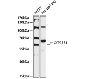
Cyp26B1 expression in A431 cells (blue line) analyzed by flow cytometry. Cells were fixed in PFA and permeabilized with 0.5% Triton. Staining was performed with Anti-Cyp26B1 Antibody (A84266) at 10µg/ml for 1 hour and Alexa Fluor 488 secondary antibody at 1µg/ml. Negative Control: Goat IgG Isotype Control (black line) followed by Alexa Fluor 488 secondary antibody.
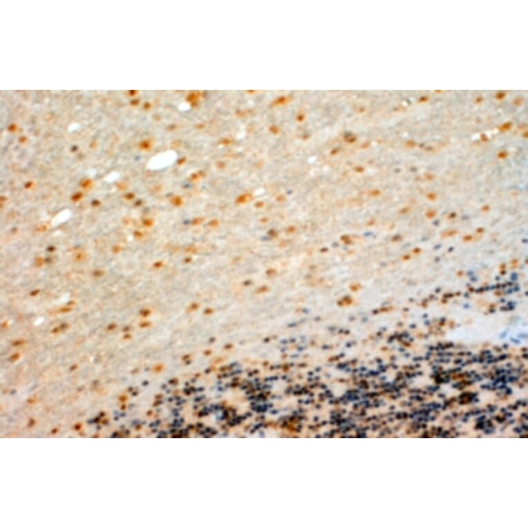
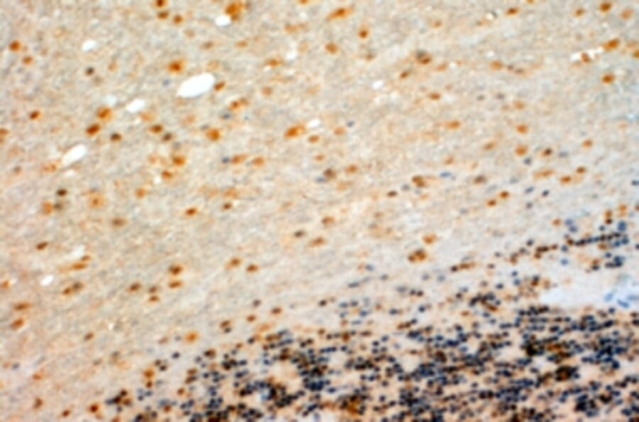
Cyp26B1 expression in Human Cerebellum analyzed by immunohistochemistry. Tissue was paraffin-embedded, and antigen retrieval was achieved by steaming in Tris/EDTA buffer, pH 9. Staining was performed with Anti-Cyp26B1 Antibody (A84266) at 4µg/ml and revealed with horseradish peroxidase (HRP).
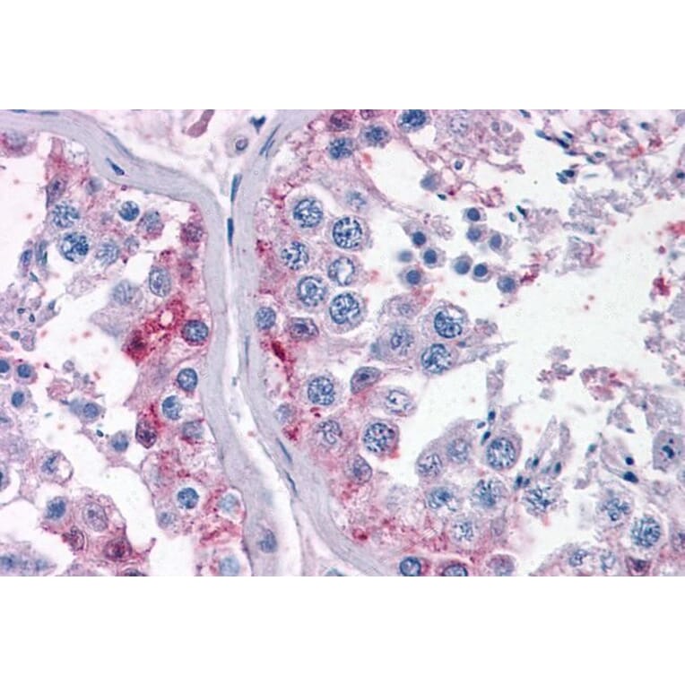
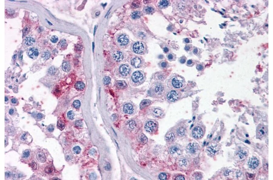
Cyp26B1 expression in Human Small Intestine analyzed by immunohistochemistry. Tissue was paraffin-embedded, and antigen retrieval was achieved by steaming in citrate buffer, pH 6. Staining was performed with Anti-Cyp26B1 Antibody (A84266) at 3.75µg/ml and revealed with alkaline phosphatase (AP).
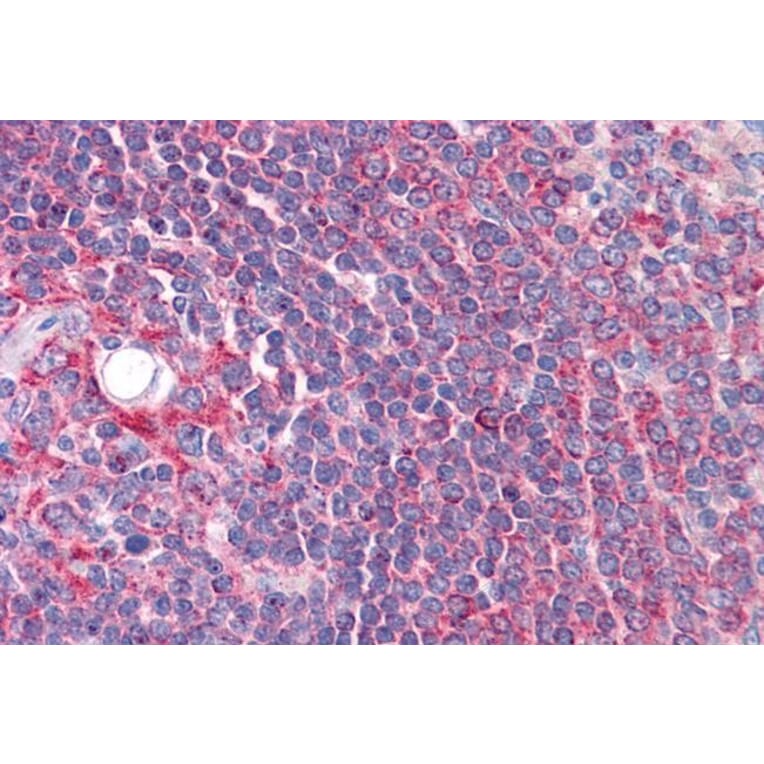
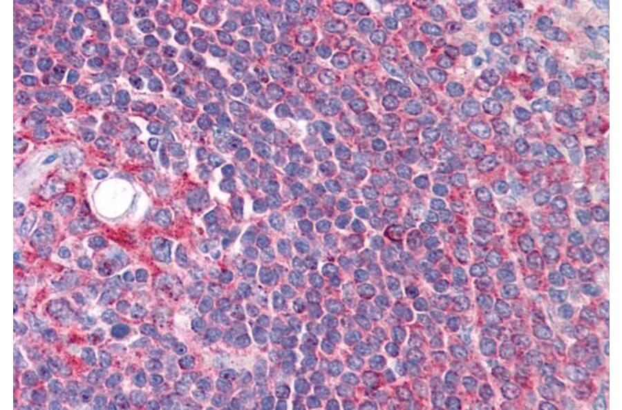
Cyp26B1 expression in Human Spleen analyzed by immunohistochemistry. Tissue was paraffin-embedded, and antigen retrieval was achieved by steaming in citrate buffer, pH 6. Staining was performed with Anti-Cyp26B1 Antibody (A84266) at 3.75µg/ml and revealed with alkaline phosphatase (AP).
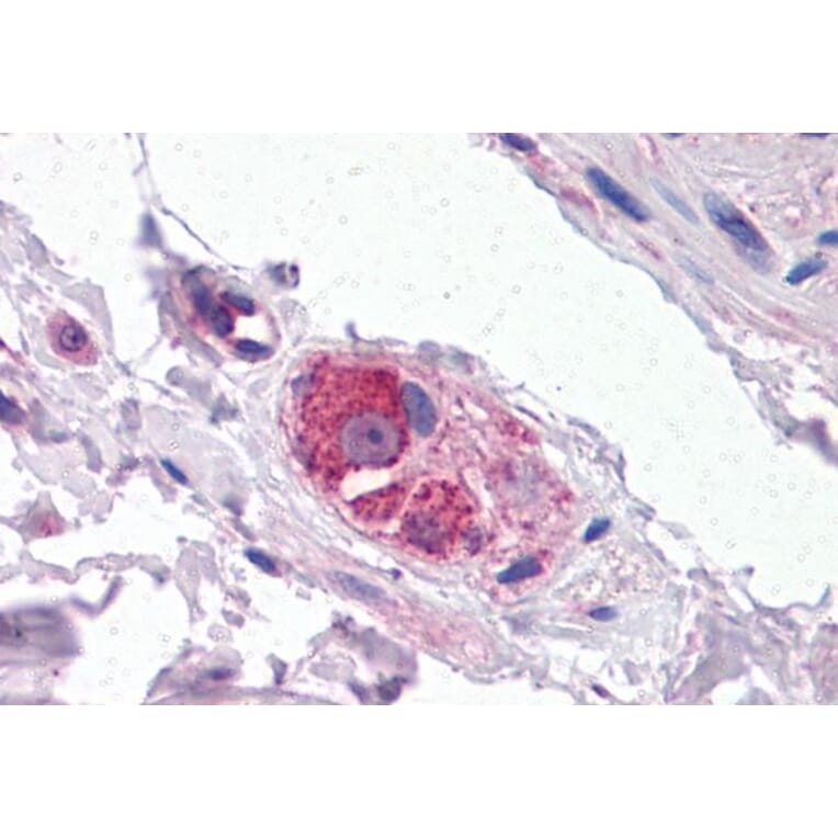
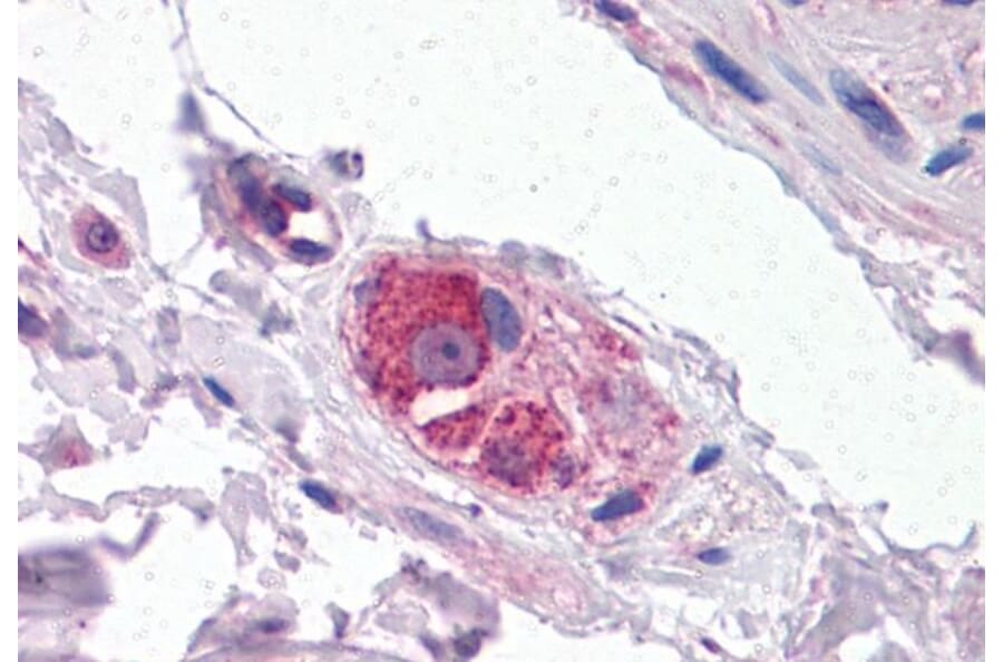
Cyp26B1 expression in Human Testis analyzed by immunohistochemistry. Tissue was paraffin-embedded, and antigen retrieval was achieved by steaming in citrate buffer, pH 6. Staining was performed with Anti-Cyp26B1 Antibody (A84266) at 3.75µg/ml and revealed with alkaline phosphatase (AP).