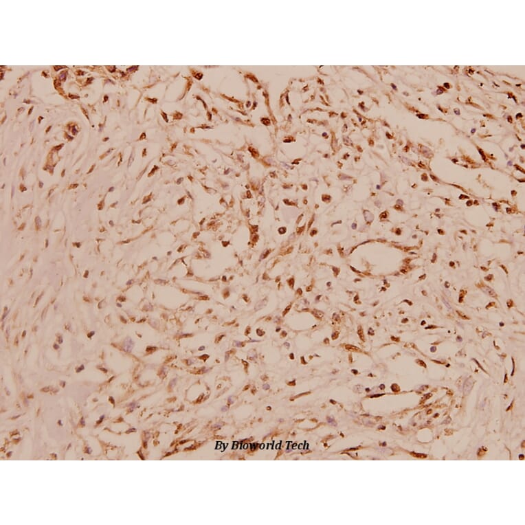
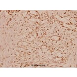
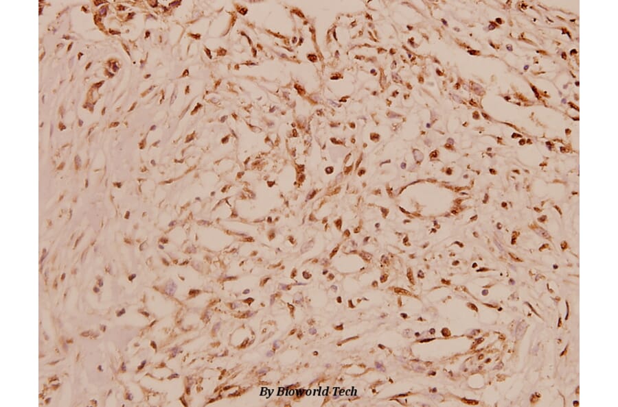
Synthetic peptide, corresponding to amino acids 400-450 of Human Cyclin A1.
Wirt
Rabbit
Klonalität
Polyclonal
Konjugat
Unconjugated
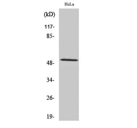
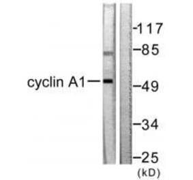
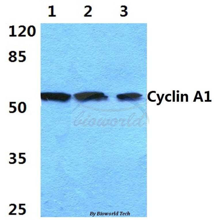
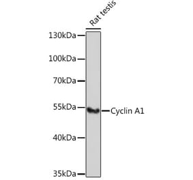
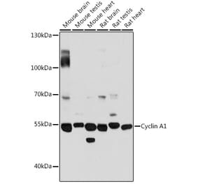
Molekulargewicht
~ 52 kDa
Reinheit
The antibody was affinity-purified from rabbit antiserum by affinity-chromatography using epitope-specific immunogen and the purity is > 95% (by SDS-PAGE).
Produktform
1 mg/ml in Phosphate buffered saline (PBS) with 0.05% sodium azide, approx. pH 7.2.


![Western Blot - Anti-Cyclin A1 Antibody [XLA1-3] (A250866) - Antibodies.com](https://cdn.antibodies.com/image/catalog/250/A250866_1.jpg?profile=product_alternative)
![Western Blot - Anti-Cyclin A1 Antibody [XLA1-3] - BSA and Azide free (A254046) - Antibodies.com](https://cdn.antibodies.com/image/catalog/254/A254046_1.jpg?profile=product_alternative)
![SDS-PAGE - Anti-Cyclin A1 Antibody [XLA1-1] (A250928) - Antibodies.com](https://cdn.antibodies.com/image/catalog/250/A250929_1.jpg?profile=product_alternative)
![SDS-PAGE - Anti-Cyclin A1 Antibody [XLA1-1] - BSA and Azide free (A254108) - Antibodies.com](https://cdn.antibodies.com/image/catalog/254/A254109_1.jpg?profile=product_alternative)