Recombinant rabbit monoclonal [DM63] antibody to CD28.
Anwendungen
ELISA, Flow Cytometry
Verdünnungen
ELISA: 1:5,000-10,000, Flow Cytometry: 1:100
Reaktivität
Human
Wirt
Rabbit
Klonalität
Monoclonal
Klon
DM63
Isotyp
IgG
Konjugat
Unconjugated
Reinigung
Affinity chromatography.
Konzentration
Reconstitution dependent.
Produktform
Lyophilized
Rekonstitution
Reconstitute with distilled sterile water.
Formulierung
Lyophilized from sterile Phosphate Buffered Saline, pH 7.4. Normally 5%–8% Trehalose is added as a protectant before lyophilization.
Lagerung
Shipped at 4°C. Lyophilized: Store at -20°C to -80°C. Reconstituted: Aliquot and store at -80°C. Product is stable for one year. Avoid freeze/thaw cycles.
Allgemeine Hinweise
Prior to reconstitution, centrifuge the vial at 5,000g for 3-5 minutes at room temperature. Reconstitute with appropriate volume of distilled sterile water to bring product to 1mg/ml concentration. After addition of distilled sterile water, mix by gentle tapping. Note: It is not recommended to vortex or vigorously pipette the sample.
Synonyme
CD 28, CD28 antigen, CD28 molecule, CD28_HUMAN, MGC138290, T cell antigen CD28, T cell specific surface glycoprotein, T cell specific surface glycoprotein CD28, T-cell-specific surface glycoprotein CD28, TP44
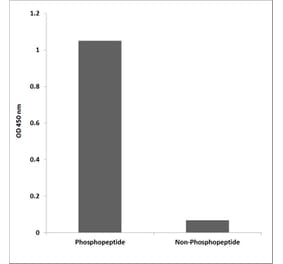
ELISA plates were pre-coated with Recombinant Human CD28 Protein (Fc Chimera 6xHis Tag) (A318394) at 2 µg/ml (100 µl/well) which can bind Anti-CD28 Antibody [DM63] - BSA and Azide free (A318634) in a linear range of 1-100 ng/ml.
Expi 293 cell line was transfected with irrelevant protein (A) and human CD28 (B) were surface stained with Anti-CD28 Antibody [DM63] - BSA and Azide free (A318634) at 1µg/ml followed by Anti-Rabbit IgG Antibody (Alexa 488).
Flow cytometry data of serially titrated Anti-CD28 Antibody [DM63] - BSA and Azide free (A318634) on Jurkat cells. The Y-axis represents the mean fluorescence intensity (MFI) while the X-axis represents the concentration of IgG used.
Affinity ranking of different Rabbit Anti-CD28 Monoclonal Antibody clones by titration of different concentrations onto Jurkat cells. The Y-axis represents the mean fluorescence intensity (MFI) while the X-axis represents the concentration of IgG used.
Flow cytometry analysis of antigen binding of Anti-CD28 Antibody [DM63] - BSA and Azide free (A318634). (A) Anti-CD28 Antibody [DM63] - BSA and Azide free (A318634) does not bind to 293T cells that do not express CD28. (B) A clear peak shift of Anti-CD28 Antibody [DM63] - BSA and Azide free (A318634) was seen compared to the control when incubated with CD28-expressing Jurkat cells, indicating strong binding of Anti-CD28 Antibody [DM63] - BSA and Azide free (A318634) to CD28. Antibodies were incubated at 2 µg/ml. .


![ELISA - Anti-CD28 Antibody [DM63] - BSA and Azide free (A318634) - Antibodies.com](https://cdn.antibodies.com/image/catalog/318/A318634_1.jpg?profile=product_top)
![Flow Cytometry - Anti-CD28 Antibody [DM63] - BSA and Azide free (A318634) - Antibodies.com](https://cdn.antibodies.com/image/catalog/318/A318634_2.jpg?profile=product_top)
![Flow Cytometry - Anti-CD28 Antibody [DM63] - BSA and Azide free (A318634) - Antibodies.com](https://cdn.antibodies.com/image/catalog/318/A318634_3.jpg?profile=product_top)
![Flow Cytometry - Anti-CD28 Antibody [DM63] - BSA and Azide free (A318634) - Antibodies.com](https://cdn.antibodies.com/image/catalog/318/A318634_4.jpg?profile=product_top)
![Flow Cytometry - Anti-CD28 Antibody [DM63] - BSA and Azide free (A318634) - Antibodies.com](https://cdn.antibodies.com/image/catalog/318/A318634_5.jpg?profile=product_top)
![ELISA - Anti-CD28 Antibody [DM63] - BSA and Azide free (A318634) - Antibodies.com](https://cdn.antibodies.com/image/catalog/318/A318634_1.jpg?profile=product_top_thumb)
![Flow Cytometry - Anti-CD28 Antibody [DM63] - BSA and Azide free (A318634) - Antibodies.com](https://cdn.antibodies.com/image/catalog/318/A318634_2.jpg?profile=product_top_thumb)
![Flow Cytometry - Anti-CD28 Antibody [DM63] - BSA and Azide free (A318634) - Antibodies.com](https://cdn.antibodies.com/image/catalog/318/A318634_3.jpg?profile=product_top_thumb)
![Flow Cytometry - Anti-CD28 Antibody [DM63] - BSA and Azide free (A318634) - Antibodies.com](https://cdn.antibodies.com/image/catalog/318/A318634_4.jpg?profile=product_top_thumb)
![Flow Cytometry - Anti-CD28 Antibody [DM63] - BSA and Azide free (A318634) - Antibodies.com](https://cdn.antibodies.com/image/catalog/318/A318634_5.jpg?profile=product_top_thumb)
![ELISA - Anti-CD28 Antibody [DM63] - BSA and Azide free (A318634) - Antibodies.com](https://cdn.antibodies.com/image/catalog/318/A318634_1.jpg?profile=product_image)
![Flow Cytometry - Anti-CD28 Antibody [DM63] - BSA and Azide free (A318634) - Antibodies.com](https://cdn.antibodies.com/image/catalog/318/A318634_2.jpg?profile=product_image)
![Flow Cytometry - Anti-CD28 Antibody [DM63] - BSA and Azide free (A318634) - Antibodies.com](https://cdn.antibodies.com/image/catalog/318/A318634_3.jpg?profile=product_image)
![Flow Cytometry - Anti-CD28 Antibody [DM63] - BSA and Azide free (A318634) - Antibodies.com](https://cdn.antibodies.com/image/catalog/318/A318634_4.jpg?profile=product_image)
![Flow Cytometry - Anti-CD28 Antibody [DM63] - BSA and Azide free (A318634) - Antibodies.com](https://cdn.antibodies.com/image/catalog/318/A318634_5.jpg?profile=product_image)
![Flow Cytometry - Anti-CD28 Antibody [CD28.2] - Low endotoxin, Azide free (A86493) - Antibodies.com](https://cdn.antibodies.com/image/catalog/86/A86496_679.jpg?profile=product_alternative)
![Immunofluorescence - Anti-CD28 Antibody [204.12] - BSA and Azide free (A253812) - Antibodies.com](https://cdn.antibodies.com/image/catalog/253/A253812_1.jpg?profile=product_alternative)
![Immunofluorescence - Anti-CD28 Antibody [204.12] (A250632) - Antibodies.com](https://cdn.antibodies.com/image/catalog/250/A250632_1.jpg?profile=product_alternative)
![Flow Cytometry - Anti-CD28 Antibody [CD28.2] (A86491) - Antibodies.com](https://cdn.antibodies.com/image/catalog/86/A86493_678.jpg?profile=product_alternative)
![ELISA - Anti-CD28 Antibody [DM64] - BSA and Azide free (A318633) - Antibodies.com](https://cdn.antibodies.com/image/catalog/318/A318633_1.jpg?profile=product_alternative)
![Immunofluorescence - Anti-CD28 Antibody [CB28] (A250629) - Antibodies.com](https://cdn.antibodies.com/image/catalog/250/A250629_1.jpg?profile=product_alternative)
![SDS-PAGE - Anti-CD28 Antibody [FR104] - Low endotoxin, Azide free (A323936) - Antibodies.com](https://cdn.antibodies.com/image/catalog/323/A323936_1.jpg?profile=product_alternative)
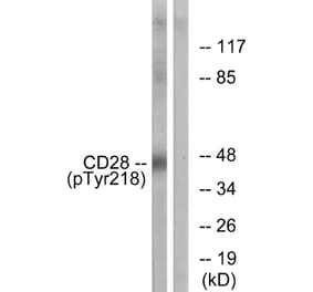
![Western Blot - Anti-CD28 Antibody [ARC5082-01] (A309309) - Antibodies.com](https://cdn.antibodies.com/image/catalog/309/A309309_1.jpg?profile=product_alternative)
![Immunofluorescence - Anti-CD28 Antibody [CB28] - BSA and Azide free (A253809) - Antibodies.com](https://cdn.antibodies.com/image/catalog/253/A253809_1.jpg?profile=product_alternative)