c-Kit expression in Human Hippocampus lysate (A) + peptide (B) analyzed by western blot. Cells were lysed in RIPA buffer and 35µg protein was run per lane. Primary antibody incubation was performed with Anti-c-Kit Antibody (A82989) at 1µg/ml and detected by chemiluminescence.
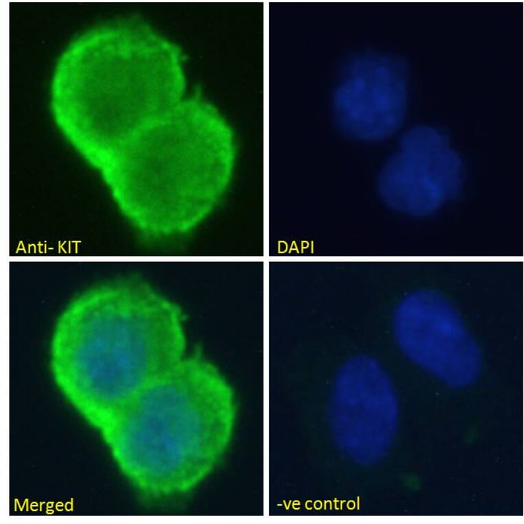
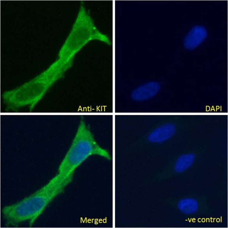
c-Kit expression in HEK293 cells analyzed by immunofluorescence. Cells were permeabilized with 0.15% Triton. Staining was performed with Anti-c-Kit Antibody (A82989) at 10µg/ml for 1 hour and Alexa Fluor 488 secondary antibody at 2µg/ml. Cytoplasmic/plasma membrane staining shown and nuclei were stained with DAPI (blue). Negative control: Goat IgG Isotype Control at 10µg/ml followed by Alexa Fluor 488 secondary antibody at 2µg/ml.
c-Kit expression in HeLa cells analyzed by immunofluorescence. Cells were permeabilized with 0.15% Triton. Staining was performed with Anti-c-Kit Antibody (A82989) at 10µg/ml for 1 hour and Alexa Fluor 488 secondary antibody at 2µg/ml. Cytoplasmic/plasma membrane staining shown and nuclei were stained with DAPI (blue). Negative control: Goat IgG Isotype Control at 10µg/ml followed by Alexa Fluor 488 secondary antibody at 2µg/ml.
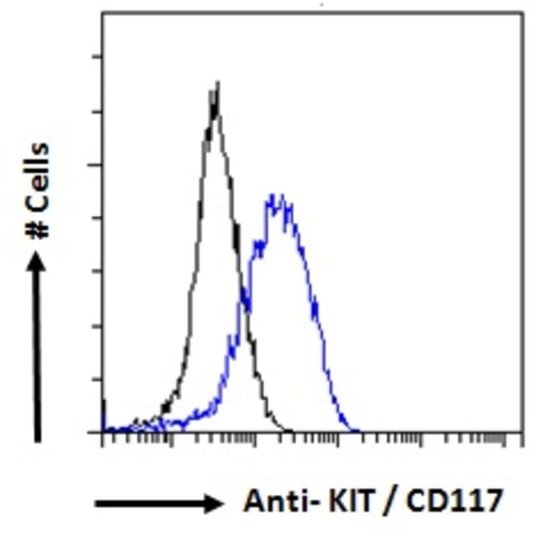
c-Kit expression in MCF7 cells (blue line) analyzed by flow cytometry. Cells were fixed in PFA and permeabilized with 0.5% Triton. Staining was performed with Anti-c-Kit Antibody (A82989) at 10µg/ml for 1 hour and Alexa Fluor 488 secondary antibody at 1µg/ml. Negative Control: Goat IgG Isotype Control (black line) followed by Alexa Fluor 488 secondary antibody.
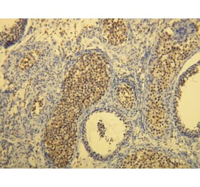
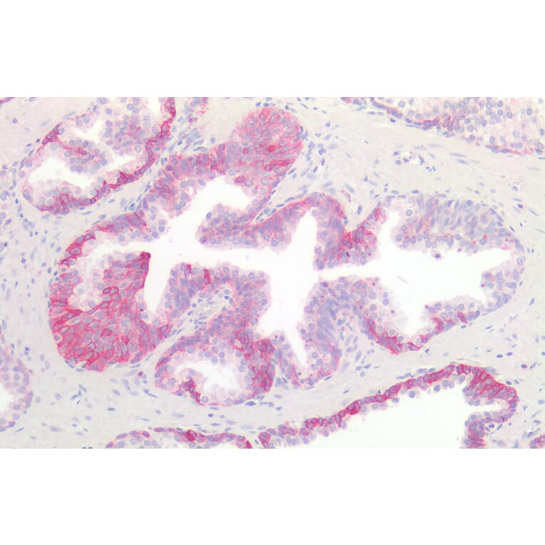
c-Kit expression in Human Prostate analyzed by immunohistochemistry. Tissue was paraffin-embedded, and antigen retrieval was achieved by steaming in citrate buffer, pH 6. Staining was performed with Anti-c-Kit Antibody (A82989) at 10µg/ml and revealed with alkaline phosphatase (AP).


![SDS-PAGE - Anti-c-Kit Antibody [LOP628] - Low endotoxin, Azide free (A323861) - Antibodies.com](https://cdn.antibodies.com/image/catalog/323/A323861_1.jpg?profile=product_alternative)
![Flow Cytometry - Anti-CD117 Antibody [104D2] (A86768) - Antibodies.com](https://cdn.antibodies.com/image/catalog/86/A86772_877.jpg?profile=product_alternative)
![SDS-PAGE - Anti-c-Kit Antibody [CDX-0158] - Low endotoxin, Azide free (A323860) - Antibodies.com](https://cdn.antibodies.com/image/catalog/323/A323860_1.jpg?profile=product_alternative)